Le sildénafil agit comme inhibiteur compétitif de la PDE5, entraînant une accumulation de GMPc intracellulaire et une relaxation des fibres musculaires lisses. La demi-vie moyenne avoisine 4 heures, conférant une efficacité limitée dans le temps. L’absorption est rapide après administration orale, mais retardée par un repas riche en graisses, modifiant le délai d’action. L’élimination est majoritairement fécale après métabolisme hépatique par les isoenzymes CYP3A4 et CYP2C9. Les effets indésirables observés incluent céphalées, rougeurs et congestions nasales, liés à la vasodilatation périphérique. Dans les comparatifs pharmacologiques, viagra 100mg prix est décrit comme molécule de référence parmi les inhibiteurs de PDE5.
Microsoft word - df front winter 2006.doc
Winter 2006
FROM THE DIRECTOR H. Leon Thacker, DVM, PhD As this is written, winter has been mild and quite pleasant compared with some of those of past years. OK with me if it continues. Activity in the ADDL continues to be high; the faculty and staff of the Laboratory continue to provide dedicated, beyond the call of duty, hustle to diagnostic requests and submissions. We have recently completed testing by the immunohistochemistry method samples from 1256 hunter-killed deer from Indiana; all samples were found to be ‘no resistant prions detected, i.e. no prions diagnostic of Chronic Wasting Disease of deer were found. We continue to support the national surveillance program for detecting Scrapie of sheep and goats. We are running 600-1000 samples by IHC per week. The federally assisted state program for
Johne’s disease surveillance in Indiana continues to generate samples for fecal culture or serum ELISA testing for Johne’s disease. Our laboratory was recently selected as one of the members of the National Animal Health Laboratory Network laboratories to participate in an interlaboratory comparison study for a newly developed Lawrence Livermore National Laboratory multiplex system capable of testing for a number of diseases from the same animal tissue/fluid sample by multiplex polymerase chain reaction methodology. High technology equipment has been installed and calibrated in the ADDL and we have two technicians in addition to the head of our molecular diagnostics area, Dr. Ramesh Vemulapalli, who have been trained on the equipment operation. I was recently presented with a letter of resignation from Dr. Zheko Kounev who has been a member of our faculty for the past three years as avian diagnostician and food safety specialist. Dr. Kounev was presented with an opportunity with an Illinois nutrition company who will assign Dr. Kounev to activity in Bulgaria for major timeframes as part of his employment responsibilities. As Zheko and his wife are natives of Bulgaria and they yet own properties there, it was an opportunity that felt they could not decline. We wish Zheko and his family the best of times in the future. ADDL will begin a search for a replacement for Dr. Kounev’s avian activities ASAP. We continue to receive queries regarding Avian Influenza presence and testing. To date, we have tested birds of several species, the most numerous being chickens and turkeys; we have found no evidence of AI presence in Indiana. Tests available in ADDL for AI include PCR, virus isolation, antigen capture ELISA and agar gel immunodiffusion. In August of 2005, we sent out a survey of ADDL laboratory users to get ideas of means whereby we can improve services. We were very well pleased with the satisfaction reflected by the returned survey results. Some areas of perceived needed improvement were identified; we are working on them. We hope to see many of the veterinarians who receive this newsletter at the annual meeting of the Indiana Veterinary Medical Association in Indy the end of this month.
FINAL DIAGNOSIS: Bone Marrow Fat Analysis as a Measure of Starvation in Animals………………………. 1 Leptospiral Reproductive Losses in Cattle…………………………………………………………………….
ADDL 2006 Schedule……………………………………………………………………………………………. 3 Equine Mandibular Juvenile Ossifying Fibromas………………………………………………………………
Granulomatous meningoencephalomyelitis (GME) in Dogs………………………………………………….
On the Road……………………………………………………………………………………………………….
ADDL News.…………………………………………………………………………………………. 7 Auto-faxing. 8 Testing for Persistently Infected BVD Animals by Antigen Capturing ELISA. 8 Antibiotic sensitivities. 9
bad teeth, parasitism, neoplasia, toxins, or
FINAL DIAGNOSIS: This column in the
Winter 2006 issue is being replaced by an
Starvation is characterized by a lengthy and
article written by Carla Vega de la Cruz,
continuous deprivation of food (Stedman’s, 1995). They both can be caused by diseases,
injuries, management conditions, and/or the
environmental conditions in which the animals
live. In the northern hemisphere, winter can
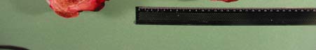
Bone Marrow Fat Analysis as a
bring on additional stress to outdoor livestock
Measure of Starvation in Animals
due to a lack of food-related negative energy
Summary: Making a definitive diagnosis of
quality/inadequate forages, cold weather, and
starvation in animals is difficult because there
increased energy demands (Radostits, 2000).
are few quantitative measures of starvation
In wild ruminants such as deer and moose,
available at postmortem examination. The
analysis of bone marrow fat content by various
Toxicology and Analytical Chemistry section of
methods has been used for several decades for
the Purdue ADDL is currently developing a
diagnosis of starvation because, following harsh
method which will be used to relate severely
winters, bones are frequently the only sample
decreased bone marrow fat to clinical starvation.
which can be found for evaluation (Cheatum,
This will be done by developing an analytical
1949, Bischoff, 1954, Greer, 1968, Neiland,
1970, Verme and Holland, 1973, Franzmann
establishing a database of values for the normal
and Arneson, 1976). In those studies, a fat
percentage of bone marrow fat in domestic
solvent extraction method was generally found
animals, and relating severely decreased bone
to provide the most consistent results when
marrow fat to clinical starvation. At this time, we
compared to other methods even though some
welcome inquiries regarding the submission of
of the other air-drying or compression methods
are more rapid and easier to perform in the field
Rationale and Significance: Malnutrition is
a state in which a diet does not provide the
Holland, 1973). In wildlife, the femur has been
optimal amount of nutrients. The long-term
effect of inadequate intake of food is starvation.
marrow fat content (Ballard, 1995). The femur is
Inadequate food intake can be exacerbated by a
used because it is readily obtained, has a large
physiological condition or disease state as well
marrow content, an abundant blood supply, and
as by extreme environmental factors such as
is one of the last fat sources to be utilized. The
those that occur in winter. Malnutrition and
bone marrow of a normal healthy animal is solid,
starvation are a natural cause of death in
white and waxy due to the high fat content
wildlife. Management practices resulting in
(Cheatum, 1949). In a state of malnutrition, the
malnutrition and starvation can also occur in
bone marrow is red, solid, and slightly fatty to
domestic livestock. When this occurs, there can
the touch (Cheatum, 1949). In an advanced
be legal ramifications related to mismanagement
state of starvation, the bone marrow is red to
and mistreatment. However, diagnostically,
yellow, gelatinous, and glistening and wet to the
touch due to the high water content (Cheatum,
starvation available at post-mortem examination.
1949). In addition to the applicability of the
This is why making the definitive diagnosis of
solvent extraction method, findings of those
starvation is many times difficult, especially if the
wildlife studies pertinent to domestic livestock
cause and time of death are unknown (Ballard,
include: 1) with a high degree of accuracy, a
1995). Therefore, a need exists for a validated,
gelatinous bone marrow ,regardless of its color,
quantitative analytical method which can be
is indicative of a poor animal resulting directly or
used to support a post-mortem diagnosis of
indirectly from malnutrition as in one study, 95%
of gelatinous marrows were found in poor deer
Literature Review: Malnutrition is defined as
and 97% were low in marrow fat (low defined in
the inadequate intake and/or malabsorption of
that study as less than 19%, Bischoff, 1954), 2)
any required nutrients (Stedman’s, 1995). This
no definite conclusion can be made from a solid
can occur in an animal which is eating, but is not
marrow concerning deer condition as in some
able to ingest, digest, absorb, and/or utilize a
sufficient quantity of nutrients (Radostits, 2000).
approximately 40%-50% fat (Bischoff, 1954), 3)
In addition to simple lack of food/nutritional
tibia marrow does not correspond to femur
intake, malnutrition can be related to injuries,
marrow (Bischoff, 1954), 4) in another study,
femur bone marrow fat content (by solvent
extraction) in winter-killed elk was less than
References:
0.25% (n=12) although the fat content in other
1) Ballard WB, Gardlner CL, Weslund JH, Miller
live elk at the end of winter could be as low as
SM: 1981. Use of mandible versus longbone to
1% (Greer, 1968), and 5) in an additional study,
evaluate percent marrow fat in moose and
femur bone marrow fat from winter-killed moose
was as low as 6.1% fat in calves and 5.5% in
2) Ballard WB: 1995. Bone marrow fat as an
adults by a dry-weight method which includes
indicator of ungulate condition-How good is it?
non-fat residue (Franzman and Arneson, 1976).
3) Bischoff AI: 1954. Limitations of the bone
performed on wildlife, there are no published
marrow technique in determining malnutrition in
reports of the use of bone marrow fat for
deer. Proc West Assoc State Game and Fish
diagnosis of starvation in domestic livestock.
(carbohydrates, protein and fat) for energy.
carbohydrate in the form of glycogen. However,
5) Franzmann AW, Arneson PD: 1976. Marrow
glycogen stores are relatively rapidly exhausted
fat in Alaskan moose femurs in relation to
and the next source for energy is predominantly
mortality factors. J Wildlife Management 40 (2):
fat. Bone marrow fat is one of the last body
stores of fat to be used. Late in the course of
starvation, when glycogen and fat stores have
indicates fat content of elk (wapiti) femur
been depleted, the only source available for
marrows. J Wildlife Management 32(4): 747-
energy is protein, the catabolism of which results
in the development of ketosis (ketone bodies in
7) Hungerford TG: 1990. Diseases of Livestock
blood and urine). If this negative energy
9th ed. McGraw-Hill Co., New York, NY. pp
balance is not corrected the animal will die. In
general, clinical signs and gross pathological
8) Neiland KA: 1970. Weight of dried marrow as
findings related to malnutrition/starvation include
indicator of fat in caribou femurs. J Wildlife
animals that are weak and underweight, have a
loss of skin turgor, have dull hair coats, sunken
9) Radostits OM, Gay CC, Blood DC, Hinchcliff
eyes, tucked-up abdomens, prominence of the
bones of shoulders, ribs, vertebra and pelvis,
Diseases of Cattle, Sheep, Pigs, Goats and
atrophy of muscles, and a decrease or absence
Horses, 9th ed. WB Saunders Co. New York,
of subcutaneous, perirenal, pericardial and bone
marrow fat which can be described as serous
10) Stedman’s Medical Dictionary. 26th ed.
Williams and Wilkins. Baltimore MD 1995.
Objective: Our goal is to develop and validate
11) Verme LJ, Holland JC: 1973. Reagent-dry
an analytical method for the quantification of
assay of marrow fat in white-tailed deer. J
bone marrow fat from the femur and utilize that
method to establish a database of normal values
in different animals for use in suspected cases of starvation. The ADDL Toxicology and Analytical Chemistry section has begun to develop and validate the analytical method. We will then develop a database of normal values that can be used to determine cases of starvation in animals. - edited by the Toxicology and Analytical Chemistry Section Dr. Steve Hooser, Section Head Dr. Robert Everson, Analytical Chemist Christina Wilson, Assistant Chemist Kim Meyerholtz, Laboratory Technician
way vaccines, but the new Spirovac vaccine for
Leptospira borgpetersenii serovar hardjo (type:
Leptospiral
hardjo-bovis) is necessary. Spirovac is safe and
Reproductive Losses
effective in calves as early as 4 weeks of age.
in Cattle
Initial vaccination includes 2 doses at 4-6 weeks
apart. Once the entire herd is initially
vaccinated, annual vaccination is required to
Traditionally, leptospiral abortions were thought
maintain protection. Additionally, any additions
of as late-term losses and the source of infection
to the herd should be isolated and follow the
was a contaminated environment due to wildlife,
same treatment and vaccination schedule used
dog, or swine reservoirs. These late-term
abortions are caused by several serovars
including Leptospira interrogans serovar hardjo
-edited by Dr. Leon Thacker, ADDL Director
(type: hardjo-prajitno), Leptospira interrogans
serovar pomona, Leptospira interrogans serovar
References:
1. ABS Global Technical Services: 2003. L.
icterohemorrhagiae, and Leptospira kirschneri
hardjo-bovis: Preventing the Silent Profitability
serovar grippotyphosa. The standard 5-way
leptospiral vaccines provide protection from all
2. Heath SE, R Johnson: 1994. Leptospirosis.
of these pathogens; however, an additional
leptospiral threat exists, Leptospira borgpetersenii serovar hardjo (type: hardjo-
Development, Infectious Disease, and Diag-
For hardjo-bovis, cattle are the maintenance
nostic Features in Cases of Pregnancy Failure.
host. The reproductive tracts and kidneys are
Proceedings Society for Theriogenology Annual
colonized and cattle serve as the source of
infection in a herd. The traditional late-term
4. Wikse SE: 2004. At Last: An Effective Control
abortions can occur, but the real effect is overall
Program for Leptospira hardjo-bovis. Veterinary
reduced reproductive performance. Herds see
5. Wikse SE: 2003. Practitioner’s Approach to
increased early embryonic losses, and stillbirths
Investigation of Abortions in Beef Cattle.
Proceedings of the Society for Theriogenology
Eliminating carriers is key to minimizing losses
Annual Conference and Symposium. 214-220.
associated with leptospirosis once a diagnosis
diagnose a herd problem with hardjo-bovis,
serum and urine samples are required from a
representative sample of the herd. Any open
cows should be included in that sampling. To
Purdue ADDL and Heeke ADDL will be closed
facilitate flushing the leptospiral organisms from
on the following University holidays in 2006.
the kidneys, cows are given furosemide and the
urine sample is collected from dilute urine that is
January 16…………….Martin Luther King Day
voided after the initial concentrated urine is
voided. The urine is chilled in red-top tubes.
July 4………………….Independence Day September 4………….Labor Day
Although serology is typically more useful with
the L. interrogans and L. kirschneri serovars,
serum samples should also be collected from
the same animals that provided the urine
samples. If aborted fetuses are available, useful
samples include kidney, liver, lung, urine, and
thoracic or abdominal fluids. Contacting your
diagnostic laboratory for guidance on what
samples to submit and how to package them for
established, carriers are eliminated by treatment
with oxytetracycline. A diligent herd vaccination
program that includes not only the traditional 5-
Equine Mandibular Juvenile
It is controversial whether the juvenile
Ossifying Fibromas
mandibular ossifying fibroma is caused by
trauma, or whether an undetected, immature
Although equine tumors are fairly uncommon, a
fibroma causes the bone to be brittle and more
significant portion of those that do arise occur in
susceptible to minor trauma. Many of the
the head and neck region. Specifically, tumors
reported cases cite an injury as the cause of the
of the oral cavity may originate in the mandible,
bony proliferation. Trauma affecting the
gums, tongue, etc., often extending into the
mandible through a fall, kick or self-inflicted
surrounding tissues. One of these tumors is the
injury (e.g., running into objects), will often result
ossifying fibroma that tends to develop from the
in gingival ulcerations and tears that will not heal
intramembranous bone of the mandible. This
tumor has a high occurrence in young (2-14
progressing to the growth of a prominent hard
structure at the site of injury. The ossifying
predilection has been shown, and genetic
fibroma arises from mutations in normal bony
predisposition has yet to be determined. Many
remodeling that would otherwise reconstruct the
believe this tumor can be diagnosed through
mandible. The mass will proliferate until the lips
history, signalment, physical examination and
are no longer apposed (allowing visualization of
radiographic findings, as it presents with highly
the mass), prehension difficulty is noted, and
characteristic features. Nevertheless, in order to
weight loss occurs as a result of not eating.
definitively diagnose an ossifying fibroma and
Many feel that the trauma sustained by the
distinguish it from similar proliferative lesions in
mandible should not be substantial enough to
the mandible (osteoma, osteosarcoma, fibrous
elicit such an injury with obvious prolonged
dysplasia, fibrous osteodystrophy), histo-
healing time. Therefore, another facet to the
pathology should be key in the diagnostic plan.
trauma theory is that the ossifying fibroma
This neoplasm is locally aggressive, with
already existed in the bone, had weakened its
extensive bony proliferation and trabecular
trabecular structure, and made the mandible
destruction, yet there are no reports of it
more susceptible to minor injury. Extensive and
possessing metastatic qualities. Medical and/or
rapid growth is then stimulated by the trauma.
surgical options may be employed to successfully treat the neoplasm, avoiding further occurrence. However, as with other invasive neoplastic processes, inadequate resection of margins or incomplete treatment often leads to rapid, extensive and increased regrowth.
Prognosis is often dependent on the extent of mandibular involvement (prehension difficulty) and aesthetic appearance of the horse (owner’s visual value). Clinical Presentation and History: Clinical
signs associated with the juvenile ossifying
Diagnosis: A tentative diagnosis can be made
fibroma depend on location and size of the
based on the history, as well as on the gross,
clinical and radiographic aspects of the lesion.
prehension, lymphadenopathy, and intermittent
Histological examination of the mass is used to
oral mucosal bleeding. Early diagnosis is not
confirm the presumptive diagnosis. This can be
often made because the anatomy of the oral
done on a core biopsy or an en bloc excisional
cavity allows for a considerable amount of
biopsy. The juvenile mandibular ossifying
involvement and progression of the ossifying
fibroma is characterized by well differentiated,
fibroma before obvious clinical signs are noted.
moderately vascularized, abundant, dense
When clinical signs are apparent, advanced
fibroblastic stroma, with isomorphic fibroblasts
local infiltration is often present. The most
transforming into osteoblasts that rim bony
common presentation is as a sub-gingival, bony
spicules. The histologic alterations tend to be
proliferation on the rostral mandible in a young
very uniform in appearance throughout the
horse. The mass is uniformly firm and does not
elicit signs of pain when manipulated. The
To distinguish the juvenile mandibular ossifying
fibroma from other closely resembling non-
mucosa covering the mass is usually ulcerated.
neoplastic and neoplastic lesions, histologic
Upon palpation, the teeth in the affected area
morphology plays an important role. Unlike
ossifying fibroma, bony spicules in fibrous
dysplasia are rarely lined by osteoblasts, and
of radiation to the tumor tissues. Radiation
only mature lesions contain deposits of lamellar
therapy can be delivered through brachytherapy
bone. Another differential diagnosis can be
or, more commonly, through an external beam.
osteoma. These are bony growths that are
External beam therapy includes gamma or X-
initially formed of cancellous bone with
rays from megavoltage equipment with Cobalt-
intertrabecular fatty or hematopoietic marrow;
they may become increasingly compact with
time. Because of the morphological similarity
treating the ossifying fibroma with a bilateral
between ossifying fibroma and some cases of
parallel opposed pair technique. The radiation
osteoma, it is thought that ossifying fibromas
margins should include the tumor and a border
of normal, healthy tissue. After several
osteosarcomas, neoplastic cells have a high
successive treatments, the mass initially
mitotic index and are pleomorphic, features
which are lacking in ossifying fibromas.
radiodense using diagnostic imaging. Over
Treatment: Treatment includes surgical
time, the ossifying fibroma progressively
(mandibulectomy, hemi-mandibulectomy) and
decreases in size to the point of no visible
Combinations of these therapies may also be
As with radiation treatment, serial follow-up
radiographs are extremely important in the
Surgical management requires the extensive
surgical post-operative monitoring of the patient.
structures (teeth), with achievement of adequate
surgery; surgery can be used to either debulk
clean margins. It has been widely reported that
the mass for radiation therapy or used in en bloc
local excision of a juvenile mandibular ossifying
excision to expose transitional margins primed
fibroma often results in rapid and proliferative
recurrence unless the surgical excision includes
In summary, mandibular juvenile ossifying
wide surgical margins. The choice of which
fibroma is a locally invasive, proliferative, fibro-
surgical procedure to use is based on diagnostic
osseous tumor that is most commonly found in
graphy), which determines the extent of bony
aggressive in nature, the neoplasm is benign, as
involvement. If diagnosed or suspected early in
no incidents of metastasis have been reported.
the growth process, a rostral mandibulectomy or
Grossly it is very distinct, yet in order to
rostral hemi-mandibulectomy may suffice as
definitively diagnose this mass, histopathology
proper treatment. If there is significant bony
involvement, more drastic surgical procedures
significant mandibular involvement, treatment
options yield a fair to good prognosis. Both
mandibulectomy) are recommended. When the
surgical and radiation therapies have resulted in
ossifying fibroma has grown from the rostral
extremely low recurrence rates when adequately
mandible, involved the entire mandibular
employed, with the horse returning to normal
prehension, activity and visual aesthetics post
mandibles, internal fixation (metal implants)
must be used to create a pseudosymphysis
upon removal of the neoplasm. This allows for
proper apposition of dentition, as well as
stabilization of the grinding forces of the jaw
during mastication. If complete removal of the
References
juvenile ossifying fibroma is achieved, there is a
very low probability of recurrence, even years
Minute Veterinary Consult-Equine. Lippincott,
post surgery. If regrowth is to occur, most
Williams and Wilkins. Iowa State University,
studies have shown that this takes place within
2. Collins JA: 1998. Ossifying fibroma/osteoma
Radiation therapy, the other therapeutic option
in the proximal tibia of a mature gelding. Vet
in cases of mandibular ossifying fibromas, uses
ionizing radiation to treat the neoplasm and to
3. Hance SR, Bertone AL: 1993. Neoplasia.
Vet Clin North Am Eq Pract. 9(1):213-234.
surpassing the normal tissue tolerance of the
4. Morse CC, Saik JE, Richardson DW, Fetter
healthy tissue surrounding the ossifying fibroma,
AW: 1988. Equine juvenile mandibular ossifying
radiation therapy delivers a sufficient lethal dose
5. Orsini JA, Baird DK, Ruggles AJ: 2004.
a) Focal GME – this is a chronic progressive
Radiotherapy of a recurrent ossifying fibroma in
condition (3-6 months) and the clinical signs
the paranasal sinus of a horse. J Am Vet Med
occur secondary to nodular granuloma formation
and mimic the effects of space occupying
6. Richardson DW, Evans LH, Tulleners EP:
1991. Rostral mandibulectomy in five horses. J
b) Multifocal or disseminated GME – This is an
acute, progressive condition (2-6 weeks). The
7. Roberts MC, Groenendyk S, Kelly WR: 1978.
most common sites affected are lower brain
Ameloblastic odontoma in a foal. Equine Vet J
stem, cervical spinal cord and meninges. Up to
25% of the dogs are dead within a week (Wong
8. Robbins SC, Arighi M, Ottewell G: 1996. The
use of megavoltage radiation to treat juvenile
c) Ocular form- this can be acute, progressive or
mandibular ossifying fibroma in a horse. Can
static and can affect eyes unilaterally or
9. www.vin.com (search juvenile ossifying
Depending on the location of the lesions, the
clinical signs can vary, but neurological deficits
common. Pathology: At necropsy, gross lesions are
evident if the angiocentric inflammation is severe and can be seen as areas of swelling and yellow
to gray discoloration. Histopathologic lesions
Please remember…
are characterized by perivascular cuffs of
cellular whorls that can evolve into nodular
granulomas (Ryan et al (Ryan et al, 2001).
Immunohistochemical characterization of the
Granulomatous meningoencephalomyelitis
inflammatory cells in the granulomatous lesions
(GME) in dogs
of GME showed that the lesions consist of MHC
class II and CD3+ T-cells indicating a T-cell
GME is an acute, progressive inflammatory
mediated delayed hypersensitivity reaction
disease of the central nervous system (CNS) of
dogs. GME is a common differential for dogs
Diagnosis: GME diagnosis is supported by the
that are affected by focal or diffuse neurological
exclusion of neoplastic, infectious and other
diseases. An inflammatory disease like GME
inflammatory conditions (e.g., canine necrotizing
can cause severe and often irreversible damage
meningoencephalitis, NME). CT and MRI can
sometimes be of use in detection of the CNS
understanding of the disease is essential.
lesions but it is difficult to differentiate the
Etiology: GME has been reported around the
lesions from neoplasia. Cell characteristics
world and can affect most breeds and ages of
such as cytologic atypia and mitotic figures
dogs; however, middle aged, small breed dogs
might be useful to differentiate this condition
from neoplasia or neoplastic reticulosis.
susceptible (Thomas, 1998). GME accounts for
Granulomatous inflammation due to viruses
up to 25% of all canine CNS disorders reported
(e.g., rabies or canine distemper), protozoa
(e.g., Toxoplasma and Neosporum), and fungi
specific etiological agent has been described for
(e.g., Cryptococcus) can be ruled out by
demonstration of specific antigens in CSF or
Clinical signs: The clinical signs of the disease
serum antibody titers for the various etiologic
are variable depending on the location of the
agents. NME can be differentiated based on
lesion in the CNS. Three syndromes of GME
breed predilection (small-size breeds, especially
have been recognized based on the location of
Treatment: B) Corticosteroids are the mainstay of treatment for GME. Response to therapy is variable and discontinuation results in recurrence of clinical signs and progression of the disease. B) Leflunomide a de novo pyrimidine synthesis inhibitor can also be used Drs. Leon Thacker, Greg Stevenson, Bob Everson, Ching
mediated) in the disease. However, these drugs
Ching Wu, Duane Murphy, Steve Hooser, Jose
are expensive and controlled clinical trial results
Ramos-Vara, Ramesh Vemulapalli, Roman
are not available. C) Radiation therapy can
Pogranichniy, and Linda Hendrickson and
prolong the mean survival (MST) of dogs.
Steve Vollmer attended the annual meeting of Prognosis: The prognosis is generally poor for
GME. MST for all dogs with GME is 14 days
Laboratory Diagnosticians in Hershey, PA,
(range 1-1215 days – Munana, 1998). Dogs
with focal signs in forebrain have an MST of
>359 days while dogs with focal signs elsewhere
Drs. Roman Pogranichniy and Ching Ching
have an MST of 59 days. Dogs with multifocal
Wu attended the Conference for Research
signs have an MST of 8 days. Dogs that receive
Workers in Animal Diseases/International PRRS
radiation therapy for focal signs can survive
>404 days. Corticosteroid therapy may induce a
transient remission of clinical signs and can
Drs. Margaret Miller, Ingrid Pardo and Gopa Gopalakrishnan attended the annual American
College of Veterinary Pathologists meeting in
Graduate Student References: 1. Cuddon PA and Smith-Maxie L: 1984.
Reticulosis of the central nervous system in the dog. Comp Cont Educ Pract Vet 6:23-32.
2. Kipar A, Baumgartner W, Vogl C, Gaedke K
and Wellman M: 1998. Immunohistochemical
characterization of inflammatory cells in brains
of dogs with granulomatous encephalomyelitis.
ADDL NEWS
3. Munana K and Lutgen P: 1998. Prognostic
Our congratulations to Dr. GopaGopalakrishnan and Dr. Alok Sharma, ADDL Graduate Students,
meningoencephalomyelitis: 42 cases (1982-
both of whom were presented awards at the recent
American College of Veterinary Pathologists
4. Ryan K, Marks SL and Kerwin SC: 2001.
Granulomatous meningoencephalomyelitis in
Gopalakrishnan was awarded one of three ACVP
dogs. Compend Contin Educ Pract Vet 23(7):
Young Investigator Awards for his poster
presentation in the Diagnostic Pathology category
5. Thomas JB: 1998. Inflammatory diseases of
entitled “Esophagitis in Camelids: Report of 3
the central nervous system in dogs. Clin Tech
Dr. Sharma was awarded the C.L. Davis DVM
Granulomatous meningoencephalomyelitis in
Foundation Student Scholarship Award in
Veterinary Pathology, an award given to a student
who displays superior knowledge of pathology as
Reporting Results
In an effort to provide results to our users
1. Ear notches should be taken with a sharp
processes to enable “AutoFaxing”. This
allows case reports to be generated, posted
to the website, and faxed shortly after the
NOT recommended; the sample they
provide is too small for an accurate test.
system, even if it is after hours or on week-
If you would prefer to have your case results
emailed instead of (or in addition to) being
samples need to be approximately 1 cm x 1 cm in size.
• Avoid testing scabby or frostbitten ears.
• DO NOT put samples in formalin.
• Samples should be submitted fresh and
days), samples can be refrigerated at 4º
From the Virology Section Testing for Persistently Infected BVD
3. Package individually in snap cap tubes (size animals by antigen capturing ELISA
the following vendors: Fisher Scientific at
Bovine viral diarrhea virus (BVDV) belongs to
the family Flaviviridae, the genus pestiviruses.
There are several members of the genus and
two types of BVD viruses. All of them are highly
• DO NOT use Whirl packs to submit
infectious and economically important to the
livestock industry. If a fetus becomes infected in utero with BVD virus in the early stages of
persistently infected (PI) without immune
response to this virus for the rest of its life. This
• It is not recommended to submit pooled
animal will shed the BVD virus in the herd and
samples for testing or to request pooling
infect other animals. If a PI animal is not
identified in the herd, it will be a source of
• If submitting more than 200 ear notches,
infection until it is removed. Elimination of BVD
from the herd requires removing PI animals from
There are several assays available to identify PI
animals: Polymerase chain reaction (PCR), virus
isolation (VI), immunohistochemistry (IHC) and
antigen capturing (Agc) ELISA. The AgcELISA
for BVD virus is a rapid diagnostic tool that can
identify PI animals in the herd if appropriate
samples are submitted. Serum and ear notch
samples are suitable for PI animal testing by
AgcELISA for BVD viral antigen. To provide
optimum service on this assay, the following
guidelines should be followed when sending ear
notch samples to Purdue ADDL for AgcELISA
Percent of Micro-organisms that are Resistence to Selected Antibiotics from Jul.- Dec. 2004 and Jan.-June 2005.
Percent of Micro-organisms that are Resistence to Selected Antibiotics from Jul.- Dec. 2004 and Jan.-June 2005.
Preoperative Instructions Plastic Surgery Pre-surgery instructions are to help reduce risks associated with surgery and anesthesia, and promote healing in the recovery period. The surgical facility or hospital will contact you before the procedure to review the preoperative instructions. Surgery may be delayed or cancelled as needed, if this pre-surgical guideline is not followed.
Gesundheit: SEXUALITÄT MITEINANDER REDEN In unserer zunehmend strukturierten und kontrollierten Welt werden freie Zeit und Genuss immer mehr zu einem Luxus. Der Einzelne ist oft überfordert. In einer solchen Zeit ist auch die Sexualität gefährdet, obwohl sie durch Nähe und Geborgenheit ein Stück Lebensqualität zurückgeben könnte. Der folgende Artikel soll Ihnen einen kurzen Einblick
 Winter 2006


 femur bone marrow fat content (by solvent
extraction) in winter-killed elk was less than
References:
femur bone marrow fat content (by solvent
extraction) in winter-killed elk was less than
References: 
 way vaccines, but the new Spirovac vaccine for
Leptospira borgpetersenii serovar hardjo (type:
Leptospiral
way vaccines, but the new Spirovac vaccine for
Leptospira borgpetersenii serovar hardjo (type:
Leptospiral 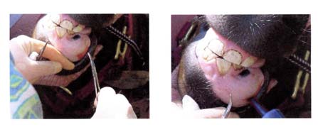 Equine Mandibular Juvenile
 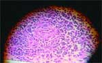 5. Orsini JA, Baird DK, Ruggles AJ: 2004.
a) Focal GME – this is a chronic progressive
Radiotherapy of a recurrent ossifying fibroma in
condition (3-6 months) and the clinical signs
the paranasal sinus of a horse. J Am Vet Med
occur secondary to nodular granuloma formation
and mimic the effects of space occupying
6. Richardson DW, Evans LH, Tulleners EP:
1991. Rostral mandibulectomy in five horses. J
b) Multifocal or disseminated GME – This is an
acute, progressive condition (2-6 weeks). The
7. Roberts MC, Groenendyk S, Kelly WR: 1978.
most common sites affected are lower brain
Ameloblastic odontoma in a foal. Equine Vet J
stem, cervical spinal cord and meninges. Up to
25% of the dogs are dead within a week (Wong
8. Robbins SC, Arighi M, Ottewell G: 1996. The
use of megavoltage radiation to treat juvenile
c) Ocular form- this can be acute, progressive or
mandibular ossifying fibroma in a horse. Can
static and can affect eyes unilaterally or
9. www.vin.com (search juvenile ossifying
Depending on the location of the lesions, the
clinical signs can vary, but neurological deficits
common.
5. Orsini JA, Baird DK, Ruggles AJ: 2004.
a) Focal GME – this is a chronic progressive
Radiotherapy of a recurrent ossifying fibroma in
condition (3-6 months) and the clinical signs
the paranasal sinus of a horse. J Am Vet Med
occur secondary to nodular granuloma formation
and mimic the effects of space occupying
6. Richardson DW, Evans LH, Tulleners EP:
1991. Rostral mandibulectomy in five horses. J
b) Multifocal or disseminated GME – This is an
acute, progressive condition (2-6 weeks). The
7. Roberts MC, Groenendyk S, Kelly WR: 1978.
most common sites affected are lower brain
Ameloblastic odontoma in a foal. Equine Vet J
stem, cervical spinal cord and meninges. Up to
25% of the dogs are dead within a week (Wong
8. Robbins SC, Arighi M, Ottewell G: 1996. The
use of megavoltage radiation to treat juvenile
c) Ocular form- this can be acute, progressive or
mandibular ossifying fibroma in a horse. Can
static and can affect eyes unilaterally or
9. www.vin.com (search juvenile ossifying
Depending on the location of the lesions, the
clinical signs can vary, but neurological deficits
common.  Reporting Results