Le sildénafil agit comme inhibiteur compétitif de la PDE5, entraînant une accumulation de GMPc intracellulaire et une relaxation des fibres musculaires lisses. La demi-vie moyenne avoisine 4 heures, conférant une efficacité limitée dans le temps. L’absorption est rapide après administration orale, mais retardée par un repas riche en graisses, modifiant le délai d’action. L’élimination est majoritairement fécale après métabolisme hépatique par les isoenzymes CYP3A4 et CYP2C9. Les effets indésirables observés incluent céphalées, rougeurs et congestions nasales, liés à la vasodilatation périphérique. Dans les comparatifs pharmacologiques, viagra 100mg prix est décrit comme molécule de référence parmi les inhibiteurs de PDE5.
Sbmj | picture quiz: cerebral abscess
studentBMJ Picture quiz: Cerebral abscess HEADLINES
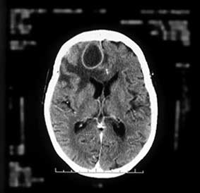
Computed tomography scan of cerebral abscess
Case history
An 82 year old woman was admitted as an emergency,
having had a generalised tonic-clonic seizure witnessed by her husband. She had no prior history of epilepsy and
no history of trauma. According to her husband she was
reasonably well and had not seen a doctor for more than
She had no history of any drug allergy and was not taking any regular medication. She was a lifelong non-smoker
Interactive
On admission she was found to have a fever, at 38oC,
and her level of consciousness was impaired (Glasgow
coma scale 6/15). Examination of her cardiorespiratory
and abdominal systems was unremarkable. She was noted to have a large boggy abscess immediately behind
Write For Us
her left ear. On further inquiry her husband acknowledged
that she had a "sore" behind her left ear that had been present for years, but his wife had never sought medical
advice. A full blood count showed a neutrophilia, but baseline investigations including a chest x ray and
electrocardiogram were unremarkable. An urgent
computed tomography scan of her brain was requested
(see figure). This was faxed through to the local neurosurgical unit which advised a course of antibiotic
therapy with repeat scan in one week. The repeat scan
was unchanged and she was subsequently transferred to
the local neurosurgical unit where she underwent stereotactic aspiration of the lesion before being
Out There
transferred back to the first admitting hospital.
http://www.studentbmj.com/issues/03/05/education/145.php (1 of 4)29/10/2005 23:07:38
Computed tomography scan of cerebral abscess
A consultant dermatologist confirmed the lesion behind her ear as a basal cell carcinoma and arranged for her to have radiotherapy. There was no evidence of sinusitis, but it was felt that the patient had a middle ear infection. She received dexamethasone and sodium valproate with blind antibiotic therapy for six weeks in total. However, blood cultures, swabs, and culture of aspirate taken during surgical aspiration were all negative. She was discharged home at seven weeks
Questions
1. What does the computed tomography scan
2. What is the diagnosis? 3. In addition to visualising the intracranial contents
on computed tomography what else might you look for on the scan?
1. Cystic lesion with a regular ring shadow with
marked surrounding oedema in the right frontal lobe.
2. Right frontal abscess (brain abscess). 3. In addition to visualising the intracranial contents,
note should also be made of the state of the paranasal sinuses and the mastoid air cells. Skull fractures and cranial defects should be looked for.
http://www.studentbmj.com/issues/03/05/education/145.php (2 of 4)29/10/2005 23:07:38
Discussion
Bacteria reach the brain via the blood-stream (for example, in congenital heart disease) and by direct extension from an adjacent focus of infection (frontal sinusitis, middle ear disease) or by implantation through wounds as a result of trauma or neurosurgery. In as many as 20% of cases the source of infection cannot be identified.
Evolution of the abscess passes through four main stages:
1. Cerebritis with surrounding oedema of white
2. The centre of the cerebritis becomes necrotic,
3. Capsule becomes more developed, a "ring"
4. Mature capsule with reactive astrocytosis.
Clinical features
Typically there is a combination of raised intracranial pressure, focal neurological signs, signs of infection, and rapid progression. Headache is common and may be diffuse, localised, and intermittent.
Emergency presentation may be with focal signs pointing to the site of the lesion, with convulsions that are usually generalised but can be focal. Up to 50% of patients may have a pyrexia. Computed tomography is the best method to confirm the diagnosis. This shows a cystic lesion with a fine but regular capsule, which enhances with contrast due to the zone of granulation tissue surrounded by oedema. If there is doubt a diagnostic burr hole and aspiration (performed stereotactically with guidance from computed tomography) is indicated.
Once the diagnosis is confirmed management will depend on several factors. If the abscess is a consequence of head trauma surgical intervention is mandatory for debridement and closure of dural defects. If the lesions are deeply seated or multiple then antibiotic treatment should be started. Close monitoring of the lesions with serial computed tomography scanning or magnetic resonance imaging is necessary and if they do not diminish in size, aspiration should be undertaken. The length of treatment is determined by clinical response and improvement of the appearance of the computed tomography scan and on average requires six or more
http://www.studentbmj.com/issues/03/05/education/145.php (3 of 4)29/10/2005 23:07:38
weeks. Dexamethasone is used to treat cerebral oedema. Rosemary Morgan, consultant, Department of Medicine for the Elderly, Arrowe Park Hospital, Upton, Wirral, Merseyside CH49 5PE studentBMJ 2003;11:131-174 May ISSN 0966-6494
http://www.studentbmj.com/issues/03/05/education/145.php (4 of 4)29/10/2005 23:07:38
Scientific Program - Tentative as on 1 January 2014 Friday 28 March, 2014 Time ST01 Urogynaecology ST02 Fetal Medicine ST03 Maternal Medicine ST04 Endoscopy ST05 RCOG International What can the RCOG do for me? Streams AM Chairs: Recurrent Urinary tract Infection: Isolated oligohydramnios in third Current guidelines for Glycemic Fertility enhancing hyster
 studentBMJ
 Computed tomography scan of cerebral abscess
A consultant dermatologist confirmed the lesion behind her ear as a basal cell carcinoma and arranged for her to have radiotherapy. There was no evidence of sinusitis, but it was felt that the patient had a middle ear infection. She received dexamethasone and sodium valproate with blind antibiotic therapy for six weeks in total. However, blood cultures, swabs, and culture of aspirate taken during surgical aspiration were all negative. She was discharged home at seven weeks
Questions
Computed tomography scan of cerebral abscess
A consultant dermatologist confirmed the lesion behind her ear as a basal cell carcinoma and arranged for her to have radiotherapy. There was no evidence of sinusitis, but it was felt that the patient had a middle ear infection. She received dexamethasone and sodium valproate with blind antibiotic therapy for six weeks in total. However, blood cultures, swabs, and culture of aspirate taken during surgical aspiration were all negative. She was discharged home at seven weeks
Questions 


 weeks. Dexamethasone is used to treat cerebral oedema.