Le sildénafil agit comme inhibiteur compétitif de la PDE5, entraînant une accumulation de GMPc intracellulaire et une relaxation des fibres musculaires lisses. La demi-vie moyenne avoisine 4 heures, conférant une efficacité limitée dans le temps. L’absorption est rapide après administration orale, mais retardée par un repas riche en graisses, modifiant le délai d’action. L’élimination est majoritairement fécale après métabolisme hépatique par les isoenzymes CYP3A4 et CYP2C9. Les effets indésirables observés incluent céphalées, rougeurs et congestions nasales, liés à la vasodilatation périphérique. Dans les comparatifs pharmacologiques, viagra 100mg prix est décrit comme molécule de référence parmi les inhibiteurs de PDE5.
S7003-.dvi
Applications of Vibrational Spectroscopy in Criminal Forensic Analysis Edward G. Bartick Handbook of Vibrational Spectroscopy
John M. Chalmers and Peter R. Griffiths (Editors)
John Wiley & Sons Ltd, Chichester, 2002
Applications of Vibrational Spectroscopy in Criminal Forensic Analysis Edward G. Bartick FBI Academy, Quantico, VA, USA This is publication number 01-06 of the Laboratory Division of the Federal Bureau of Investigation. Names of commercialmanufacturers are provided for identification only and inclusion does not imply endorsement by the Federal Bureau ofInvestigation.INTRODUCTION TO FORENSIC
Individual characteristics are properties of evidence that canbe attributed to a common source with an extremely high
ANALYSIS
degree of certainty. Class characteristics are properties ofevidence that can only be associated with a group and never
Sir Arthur Conan Doyle is believed by many to have first
popularized the application of forensic analysis through hisfictional character Sherlock Holmes, originally published
Fingerprint and DNA evidence are accepted as having
in 1887. This work is thought to have inspired many of
individual characteristics. However, fibers or copy toners
the early forensic scientists. One of these was Frenchman
are identified by their class characteristics and, because
Edmond Locard, who proposed that when two objects come
of the large production of these materials, they cannot be
into contact with one another, a cross-transfer of evidence
individualized. The strength of fiber evidence depends on
occurs.1,2 This statement is known as Locard’s Exchange
the likelihood of those same type of fibers being randomly
Principle and is the foundation for use of physical evidence
located on the suspect. Common fibers such as blue or white
to link or at least associate a suspect to a crime scene or
cotton from jeans or shirts, respectively, have little eviden-
a victim. Depending on the nature of the evidence, a wide
tial value. But blue nylon-6,6 fibers with an unusual cross-
range of analytical methods are used in forensic casework.
section would have more significant value, because there
This article illustrates how vibrational spectroscopy is used
are fewer in existence. Forensic scientists have thoroughly
to identify or compare physical evidence in criminal foren-
developed statistical values for DNA and fingerprint data to
demonstrate individual characteristics. Statistics are more
A broad definition of the term “forensic”, according
difficult to apply to class evidence, but approaches to apply
to ‘Webster’s New World Dictionary,’ is “suitable for
them are being investigated. The information obtained by
a law court or public debate”. With the application of
vibrational spectroscopy is usually characteristic of classes
forensic science, one must demonstrate in court that the
of materials, but in some instances the identification of spe-
evidence analyzed has relevance to the case in question.
cific components demonstrates an uncommon characteristic.
The significance of evidence related to a case is often
If a contaminant on an evidential material is identified as
determined by whether the physical evidence has individual
a rare substance specific to the crime scene, the evidence
would demonstrate a high likelihood of originating fromthe crime scene. Therefore, vibrational spectroscopy is used
This is a US Government Work and is in the public domain
to identify chemical properties that contribute in varying
degrees to the evidential value in criminal forensic analysis. Forensic Applications of Vibrational SpectroscopyGENERAL USE OF VIBRATIONAL
sample preparation, they are used at least as frequently as
SPECTROSCOPY IN FORENSIC
Gas chromatography (GC) with IR detection (GC/IR)
ANALYSIS
for analysis of mixtures has not found much applicationin forensic analysis. GC combined with mass spectrome-
Infrared spectroscopy
try (MS), or GC/MS, superseded GC/IR, and the ultimatesensitivity benefits of MS for trace components have over-
Several authors have provided general overviews for the use
shadowed the use of GC/IR in forensic analysis. However,
of infrared (IR) spectroscopy in forensic analysis.4–6 The
GC/IR used as a separation and identification tool for large
applications vary to cover a wide range of physical evidence
samples has proven successful in drug analysis. Because IR
in the form of bulk materials and micro-sized particles. His-
analysis can be used to identify isomers, it can sometimes
torically, dispersive IR spectrometers were generally found
be used to identify isomeric forms that render a drug active
useful for bulk samples such as drugs. Micro-sized sam-
ples were analyzed with skilled patience in beam condenser
The recent developments of single or multiple reflection,
accessories. For example, the Royal Canadian Mounted
horizontal ATR accessories for use in sample compartments
Police (RCMP) Forensic Service Laboratories (FSL) used
have been very useful for forensic analysis. Small liquid
high-pressure diamond-anvil cells in beam condensers for
or solid samples, less than a millimeter in diameter, can
the analysis of automotive paint chips transferred from hit-
be measured. Typically, diamond internal reflection ele-
ments (IREs) are used for durability. Some IREs consist
With the development of Fourier transform infrared
of thin diamonds backed with other materials to reduce
(FT-IR) spectroscopy, the application of IR to forensic
cost. These ATR accessories offer the option of extended
analysis became more prevalent because of the increased
frequency ranges to near 200 cm 1 in spectrometers with
speed and sensitivity of FT-IR. The development of dif-
cesium iodide optics. The extended range can be used to
fuse reflection (DR) accessories provided ease of sample
nondestructively acquire information regarding paint pig-
introduction for several forensic applications. Samples with
ments and inorganic fillers in polymeric materials.
matte-finished surfaces could be analyzed with no samplepreparation. Samples such as illicit drugs that previouslyrequired extensive grinding to make KBr pellets required
Raman spectroscopy
less preparation. Suzuki was the first to apply the DRmethod to forensics with the analysis of drugs, polymers,
Recent technological advancement in Raman spectrometers
wood and solvents.10 He continued with additional work on
has provided a reason for exploring this method in foren-
drugs,11 polymer foams12 and paints.13,14 Document analy-
sic applications. While the applications have been slow to
sis by DR has been reported for copy toners15,16 and inks.17
find their way into forensic laboratories, the advantages
It was not until the 1990s that the use of FT-IR became
are being recognized and Raman spectrometers are start-
more regularly applied in forensic laboratories. The intro-
ing to find use in forensic analysis.20 Fourier transform
duction of lower cost spectrometers and microscopes paved
(FT) Raman received early attention in the redevelopment
the way for FT-IR use in forensic analysis. FT-IR micro-
of Raman technology. The 1064 nm near-infrared (NIR)
scope development is considered a milestone achievement
excitation laser that is used with FT systems causes fewer
for forensic analysis and is considered the most signifi-
samples to fluoresce than the visible wavelength lasers
cant recent advancement for microsample analysis.5 Sample
previously used. However, with the lower energy, longer
preparation to introduce specimens into microscopes is fre-
wavelength laser, signal averaging and rapid scans pro-
quently easier, because only a small portion of the sample
vided by FT systems are required to attain the quality of
is required for placement in the IR beam for transmis-
spectra desired. Unfortunately, the lower scattering power
sion spectroscopy. For example, to analyze paint from a
of the longer wavelength excitation impedes application
surface, all that is required is a sliver sliced from the
to microsamples that are commonly required with foren-
surface with a scalpel. Therefore, standard sized samples
sic analysis, thus limiting the use of microscopes with FT
are often reduced in size and analyzed in microscopes
Raman. Dispersive Raman systems that use NIR lasers
because of convenient sample preparation.18 The reflection
ranging from 780 to 840 nm have greater scattering effi-
techniques, reflection-absorption (R-A), specular reflection,
ciency. Combined with sensitive charge coupled device
diffuse reflection (DR), and internal reflection spectroscopy
(CCD) detectors and improved monochromators, these sys-
(IRS), frequently referred to as attenuated total reflection
tems have more general use compared to FT Raman systems
(ATR),19 provide additional ease of sampling in IR micro-
and have thus taken the forefront for a wide range of
scopes. Because reflection methods require little or no
sample analysis. Additionally, fiber optic probes are used
Applications of Vibrational Spectroscopy in Criminal Forensic Analysis
on dispersive systems to sample through glass bottles and
plastic bags, making noninvasive sampling possible. Theseprobes contribute to the application of small, rugged Ramanspectrometers designed specifically for field use. Portabil-ity and ease of sampling are attractive features that areincluded in these Raman systems that can be used alongwith an array of other analytical instruments to be carried
to crime scenes where the analysis of potentially hazardousmaterials is conducted without risk of transporting them
POLYMER ANALYSIS General polymers
Vibrational spectroscopy is applicable to a wide range of
physical evidence. Because polymers are so common, they
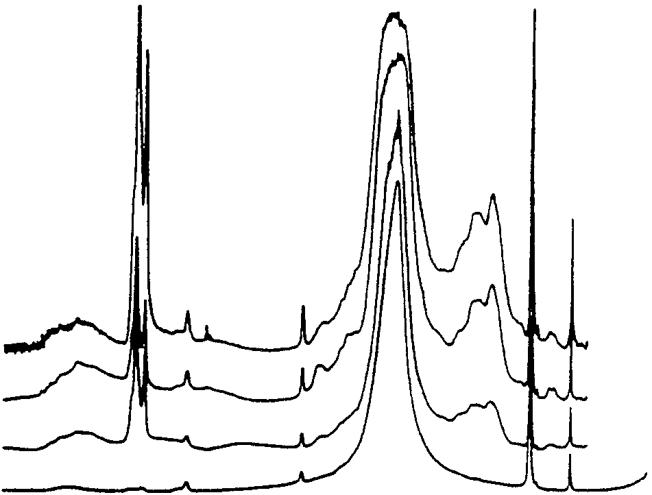
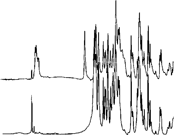
Figure 1. Cruise ship homicide case. Spectra of rubber particle
frequently play an evidentiary role in criminal cases. Poly-
evidence from sweat pants from both (a) the victim and (b) the
meric materials such as fibers, paints and adhesive tapes are
suspect, (c) running track and (d) calcite reference.
frequently analyzed to identify characteristic information
Particles from the track material were prepared in the same
regarding their composition. Physical and chemical infor-
way, and the spectra of all three samples were compared
mation on these materials is stored in computer databases
as shown in Figure 1. The three spectra of the rubber-
to help determine the manufacturer or, supplier, or simply
ized material matched closely. The material was heavily
to discriminate between many similar samples of material.
Some of the available databases will be described as part
C–O antisymmetric stretch near 1450 cm 1, and narrow
of the analyses mentioned in the forthcoming pages. Other
out-of-plane and in-plane bends near 880 and 710 cm 1,
general polymeric materials found as evidence do not fall
respectively.21 To determine the force required to embed
into a particular category and must be studied on a case-
the particles in the pants, a fabric similar to that of the
by-case basis without the aid of comparison with similar
pants was rubbed across the removed track piece at differ-
ent degrees of pressure. The investigators found that it was
The following is a case example where a polymeric mate-
necessary to rub the fabric with significant pressure, such as
rial became important evidence. Early one morning, on a
in the case of dragging a person, to cause the transfer and
luxury cruise ship off the coast of California, a man reported
embed the material in the fabric. They determined that the
to the captain that his wife had been blown overboard. Con-
transfer of particles suggested an altercation between the
sidering the minimal wind conditions, the captain became
individuals, and, therefore, this evidence weighed heavily
suspicious and alerted the law enforcement authorities. Sev-
and resulted in a homicide conviction. The combined com-
eral hours after the overboard report, the woman’s body
parison of evidential materials by microscopic examination,
was recovered by the US Coast Guard. The clothing that
IR analysis, and physical testing played a significant role
both the man and woman were wearing at the time of
the incident was forwarded to the FBI Laboratory. Thewoman’s running pants appeared soiled with an orangesubstance on both legs. Careful examination of her pants
Copy toners
with a stereo microscope revealed orange-colored, rubberyparticles. Examination of the man’s running pants also
Questioned documents involving fraud and threatening let-
revealed rubbery particles that appeared identical. On the
ters are often produced on printers, copy machines and
ship’s deck, at the location of the overboard incident, there
facsimile machines. The machine model identification of
was an orange-colored, rubberized running track. A por-
this common office equipment has been achieved through
tion of the track material was removed from the ship and
comparison of the resins of the toners used as ink. These
forwarded to the laboratory for analysis. The particles were
“copy toners” have been studied for forensic analysis as
removed from both pairs of running pants and flattened in a
a class of polymeric material. An example where copy
compression cell to make them sufficiently thin to perform
toner analysis was used to produce an investigative lead
transmission analysis using an FT-IR microscope system.
was in a case involving a copied address label. A packaged
Forensic Applications of Vibrational Spectroscopy
bomb, mailed to a corporate executive, had an address label
baseline flattened spectrum in Figure 2(b) is typical of a
that appeared to be an enlarged copy of the company’s
styrene/acrylate copolymer. Significant variations in the IR
return address logo typically used on company envelopes.
spectra are produced by the polymeric resins which contain
Investigators suspected that the bomb had been mailed by
numerous additives that vary in type and quantity. A visual
an employee with access to internal supplies and that the
comparison of the case sample spectrum was made with the
person had copied an envelope using equipment within the
62 spectra of the model types in the building. One Kodak
company. There were over 200 copy machines, involving
model type matched closely with the case sample spec-
62 different copier models, located throughout the facil-
trum. The spectrum was also searched in an IR database
ity. It was important for the investigators to know the
of copy toner resins categorized based on over 800 copier
copier model used to narrow the area of the investigation to
and printer models.23 The search software narrowed the
employees with convenient access to a copier model of the
toner type to a group containing 24 models of machines.
type used to print the label. Sample pages were prepared
By careful scrutiny of the peaks, it was possible to narrow
from each of the 62 models and forwarded to the FBI Lab-
the spectra to six Kodak models in the database. The Kodak
oratory for analysis. In the laboratory, the samples were
copier model from the corporate building was included in
prepared for IR analysis using a heat transfer technique
the computer search. Therefore, the results of the visual
to remove the toner from the documents. The preparation
inspection and the computer search of the spectra corrob-
technique involves heating the back of the paper with a
orated. Two-thirds of the binders contained in the spectral
soldering iron at a specified temperature and smearing the
database consist of the styrene resins plus additives to pro-
toner onto aluminum foil attached to a glass microscope
vide desired properties in particular copy machines. Other
slide. Spectra were obtained with an FT-IR microscope
types of binders used are phenolic and polyethylene resins.
by R-A. With this method, the IR beam passes through
The regions boxed off in Figure 2(b) contain small bands
the sample and is reflected from the aluminum foil to the
from the additives that provide the differentiating spectral
detector via the microscope optics.22 Figure 2(a) shows
features of this toner resin. Because the building contained
the original spectrum of the toner from the bomb pack-
only eight examples of that particular Kodak model, the
age label. This spectrum is sloped due to scattering from
results of this analysis permitted the investigators to narrow
the carbon black particles used for the copy image. The
their search to personnel working in limited locations of the
Figure 2. Poly(styrene:acrylate) resin copy toner spectrum from an address label on a bomb package: (a) original uncorrected spectrum; (b) flattened spectrum showing boxed regions where additive absorptions can be observed. Applications of Vibrational Spectroscopy in Criminal Forensic Analysis
building. Thus, a suspect was determined in considerably
position or environment within the comonomer structure as
less time than if the company’s entire personnel required
a methacrylate, methyl methacrylate or vinyl acetate. Over
20 variations of acrylics can be identified by IR.30 Thus, IRis a very useful tool in providing information that furtherdiscriminates fiber types to enhance the evidential value of
Raman spectrometry promises to complement IR analysis
Textile fibers are often transferred between clothing dur-
of textile fibers due to the ease of sampling and the addi-
ing personal contact in violent crimes such as rape andhomicide. The foremost method of analysis for this type
tional information it provides. Light microscopy analysis
of evidence is visual light microscopy, though IR is very
requires fiber samples to be mounted in a liquid medium,
useful to further specify fiber type. Fibers from the clothing
under coverslips, on a glass slide. Because glass absorbs
of the victim and suspect are screened microscopically for
strongly in the IR, the fibers must be removed and cleaned
similarity, closely comparing physical and optical proper-
prior to IR analysis. Raman analysis, on the other hand,
ties of the fibers. These properties may vary significantly
has been successfully performed on single fibers mounted
due to the color, shape, texture, and chemistry of textiles.
on glass microscope slides,31,32 thus avoiding the need to
Polarized light microscopy is used to determine the generic
remove the fibers from the slide mount. The additional sam-
classification of the polymer type, and IR microscopic anal-
ple preparation time is not required and, once mounted,
ysis plays an important role by identifying subclasses of
the chance of fiber loss is minimal. Figure 4 illustrates
synthetic fibers.24,25 A spectral library of 83 polymeric fiber
the process of obtaining a nylon-6 fiber spectrum using
types, obtained by transmission spectroscopy on flattened
microscopic Raman analysis. Figure 4(a) includes spectral
single fibers, was developed to aid forensic examiners in
contributions from the fiber, Permount mounting medium
the identification of fiber composition.26–29 IR is particu-
and glass from the coverslip. Figure 4(b) shows the result
larly useful for subclassifying acrylic fibers that are seen
of Permount subtracted from the original spectrum. The
frequently as a wool alternative in sweaters and readily
nylon-6 spectrum remains in Figure 4(c) after the glass
transfer between individuals during contact. These fibers
contributions are subtracted. The baseline is flattened for
consist of at least 85% polyacrylonitrile28 plus copoly-
the final spectrum shown in Figure 4(d). The signal-to-
mers and ionic end-groups to enhance dyeing properties.
noise ratio is less than obtaining a spectrum without being
Figure 3 shows spectra of the three most common acrylic
mounted under a coverslip, but it is sufficient to identify
copolymers. The band shape of the C–O stretch in the
the fiber by the prominent characteristic bands as labeled.
region between 1300 and 1000 cm 1 varies depending on its
The band assignments of the major peaks are labeled in
Figure 3. IR spectra of acrylic copolymer fibers: (a) poly(acrylonitrile:vinyl acetate); (b) poly(acrylonitrile:methyl acrylate); (c) poly(acrylonitrile:methyl methacrylate). Forensic Applications of Vibrational SpectroscopyFigure 4. Raman spectra of a nylon-6 fiber mounted under a coverslip on a glass microscope slide: (a) original spectrum, (b) Permount mounting medium subtracted; (c) glass subtracted; (d) baseline flattened with the major characteristic bands labeled.
accordance with Hendra et al.33 Raman spectra differ from
IR spectra because the selection rules for Raman vary fromIR, thereby producing complementary information. The
IR spectroscopy of paints has been useful in forensic analy-sis since the 1960s. Automotive, architectural, art, marine,
information obtained by Raman is at times more definitive
aircraft, tool, and other types of paints may become evi-
in determining the polymeric structure. Since Raman spec-
dence in a variety of crime scene scenarios.36 After visual
troscopy demonstrates spectral response from dyes, the
light microscopy, IR analysis offers the most information
dye information can be useful. However, the dye spectral
in forensic paint examination. The organic binders are fre-
features can interfere with identification of the polymeric
quently identified with IR, and both organic and inorganic
composition of fibers. A protocol to determine whether to
pigments can often be identified. Scientists from the RCMP
use Raman or IR when analyzing dyed and undyed fibers,
have been classifying automotive paints based on chemical
grouped by generic class, is currently being established
composition since the 1970s.7–9 The original analysis was
in the FBI Laboratory. To further characterize the fibers,
performed with the use of high-pressure diamond-anvil cells
dye spectral features could provide information regarding
in beam condensers on dispersive IR spectrometers. Since
the dye type. Studies have been conducted using surface
then, the RCMP and other analysts have changed to using
enhanced Raman spectroscopy (SERS) to study dye compo-
the less cumbersome low-pressure compression diamond
sition using silver colloid substrates.34,35 While the spectral
cells with beam condensers in FT-IR systems. Inorganic
features are significantly enhanced by SERS, this method
pigment components in paints have revealing spectral fea-
requires that the fibers are removed from the glass slides,
tures at the lower wavenumbers. Beam condensers are
resulting in an additional step and a chance of fiber loss.
used rather than FT-IR microscopes to overcome the lim-
Raman analysis has demonstrated certain advantages over
ited frequency range of mercury cadmium telluride (MCT)
IR, but the strengths and limitations of both are still under
detectors used in IR microscopes. For paint analysis, the
study to determine where each of the methods can be used
extended range to near 200 cm 1 is obtained with CsI optics
to obtain the greatest information with the most convenient
and a standard deuterated triglycine sulfate (DTGS) detector
Applications of Vibrational Spectroscopy in Criminal Forensic Analysis
Physical and chemical information obtained by light
Researchers are beginning to demonstrate the usefulness
microscopy and IR analysis on automotive paint, used to
of Raman analysis for organic and inorganic pigment
potentially identify make, model and year of vehicles, is
identification in paint.38,39 Figure 5 compares IR and
readily searched in a database. The computerized library,
Raman paint spectra of a yellow acrylic melamine enamel
developed by the RCMP, is widely used by forensic lab-
automotive paint. The IR spectrum in Figure 5(a) clearly
oratories throughout North America.37 Necessitated by the
shows the resin binder features. The N–H stretch near
international nature of the automotive industry, European,
3350 cm 1, the C–H stretches near 3000 cm 1, the CDO
Japanese and Australian forensic laboratories will soon be
stretch near 1730 cm 1, the C–N stretch near 1540 cm 1,
contributing to this database in order to provide a more
and the typical C–O envelope from 1300 to 1000 cm 1 are
comprehensive collection. Scientists from forensic working
observed in the IR. Of particular interest to paint analysis
groups of the listed countries are contributing automotive
are any contributions by pigments. The weak, broad band at
paint samples and data from their respective nations and
868 cm 1 appears to be contributed by chrome yellow, as
shown in the reference spectrum (Figure 5(b)). However,
Paint types other than automotive do not usually dis-
because of the band’s comparatively low intensity and lack
play as much diversity in chemistry, color and layer
of detail in the paint spectrum, it would be difficult to
structure. Therefore, a wider variety of analytical instru-
positively identify chrome yellow by this method alone.
ments are often used to characterize these paints. Along
The peaks labeled at 659, 425 and 357 cm 1 are rutile,
with microscopy and IR spectroscopy, other methods fre-
a crystal form of titanium dioxide. Figure 5(d) is the
quently used are pyrolysis GC/MS and inorganic analysis
Raman spectrum of the yellow auto paint. The major
by scanning electron microscopy with energy dispersive X-
peaks at 843 and 365 cm 1 match up with the Raman
ray spectroscopy (SEM/EDX) or X-ray diffraction (XRD)
spectrum of chrome yellow shown in Figure 5(c). Peaks
at 611 and 446 cm 1 are contributed by rutile, as shown in
3600 3200 2800 2400 2000 1800 1600 1400 1200 1000
Figure 5. IR and Raman spectra of yellow acrylic melamine enamel auto paint with pigments: (a) IR spectrum of the auto paint; (b) IR spectrum of chrome yellow pigment; (c) Raman spectrum of chrome yellow pigment; (d) Raman spectrum of the yellow auto paint; (e) Raman spectrum of rutile. (Spectra provided by E. Suzuki, Washington State Patrol, Forensic Laboratory, Seattle, WA.) Forensic Applications of Vibrational Spectroscopy
Figure 5(e). With Raman, the high scattering efficiency of
permits acquisition of spectral features of the inorganic
some pigments, relative to those of binders, helps to easily
fillers commonly found in duct tape adhesives and less
determine the pigment components, as interfering binder
frequently found in the film backing. Figure 6(a) shows
an atypical duct tape backing spectrum containing cal-cium carbonate (calcite). The calcite filler has a latticeband21 near 315 cm 1 that would not have been observed
without the extended frequency range capabilities. The C–Oasymmetric stretching band21 near 1450 cm 1 underlies the
Pressure-sensitive adhesive tapes play an important role in
C–H bending band near 1460 cm 1. The C–O out-of-plane
forensic analysis as evidenced by their diverse uses. Elec-
bend can be observed near 880 cm 1. The spectrum in
trical tapes can be used in wiring electronic devices to
Figure 6(b) is more typical of polyethylene backing show-
bombs, duct tapes in binding victims of violent crimes, and
ing only the C–H bands without the filler features. Because
other tapes in wrapping packages containing drugs, bombs
fillers are not typically used in the backings, analysis of
or other threatening material. By carefully characterizing
this unusual duct tape evidence could provide increased
tapes submitted as evidence, they can be compared with
known tapes in a suspect’s possession or they can be stud-
To more fully characterize tapes, other analytical meth-
ied to develop investigative leads when the brand can be
ods are used. The physical characteristics are observed and
established. IR spectroscopy can be useful with ATR acces-
measured with the unaided eye and microscopically, and
sories in determining the major organic components of both
inorganic composition is determined by SEM/EDX and
the adhesives and backings. In the past multiple reflection
XRD analysis. Physical characteristics, such as the yarn
accessories were used, but more recently single reflection
counts and weave type on the fabric reinforcement within
ATR accessories for the IR microscopes have been used
duct tapes, may quickly narrow down the brand possi-
to acquire spectra of small, uncontaminated areas on the
bilities. XRD can provide further information about the
tapes. While this is convenient, the frequency range is lim-
inorganic components like distinguishing between anatase
ited by the MCT detectors used on the microscopes which
and rutile crystalline forms of titanium dioxide extenders.
cut off near 700–650 cm 1. A study has demonstrated the
TiO2 is frequently used in duct tape adhesives and some
usefulness of a single-reflection, horizontal sample com-
manufacturers prefer a specific crystalline structure, thus
partment accessory using a diamond interface backed with
identification of the mineral type can assist with identify-
KRS-5.40 This accessory provides a spectral range from
ing the manufacturer. All of the information obtained by the
4000 to 260 cm 1when used with cesium iodide optics
various analyses has recently been placed in a searchable
in the spectrometer. The extended range below 400 cm 1
database for quick comparison of tape properties.41
Figure 6. IR spectra of duct tape backing film by ATR: (a) typical polyethylene backing; (b) backing containing calcite. Applications of Vibrational Spectroscopy in Criminal Forensic Analysis
IR has been used for the analysis of both licit and illicit
drugs for many years.4,42 The computerized drug libraryproduced at the Georgia State Crime Laboratory (GSCL)
is a standard in forensic analysis. Currently, it containsover 2000 spectra of drugs and related chemicals. Samples
Figure 8. Reduction reaction of ephedrine to methamphetamine.
prepared in standard 13-mm KBr pellets have been used forinclusion of drug spectra in the library. However, recently
stereoisomers (d and l forms). IR is more effective than MS
the GSCL successfully applied ATR to drug analysis.43
in differentiating diastereoisomers. Thus, the diastereoiso-
Horizontal ATR sample compartment accessories with three
mers ephedrine and pseudoephedrine, which are precur-
reflections provide sufficient sensitivity to acquire spectra of
sors for methamphetamine, are identifiable compounds by
approximately 400 ng of lysergic acid diethylamide (LSD)
this method. Figure 8 shows the reduction reaction of
as a film cast from chloroform (Figure 7). The region
the ephedrine to methamphetamine. Figure 9 illustrates
between 2400 and 1800 cm 1 was blanked to remove the
the comparison of the gas phase spectra of these com-
uncompensated diamond absorption produced by the IRE.
pounds. In Figure 9(a) and (b), the region between 1300
The ATR spectra of drugs can be successfully searched
and 1000 cm 1 shows subtle but consistently different spec-
in the original transmission spectral library in spite of the
tral features of the diastereoisomers ephedrine and pseu-
intensity differences in the peaks. This method is beginning
doephedrine. The spectra of amphetamine and metham-
to gain acceptance in laboratories around the USA. The
phetamine, shown in Figure 9(c) and (d), respectively,
FBI Laboratory has started an ATR database of drugs for
do not have the OH stretching band near 3600 cm 1,
computer spectral searching. Recently, a drug library of 455
because the reduction reaction removes the OH attached
spectra by ATR was produced at the Illinois State Police
to the carbon next to the phenyl group. The spectra of
amphetamine and methamphetamine differ in the entire
GC combined with IR (GC/IR) simplifies the analy-
region below 1700 cm 1. The most pronounced difference
sis of drug mixture samples typical of those associated
lies with the NH deformation band near 1600 cm 1, which
with clandestine laboratories and is a standard procedure
is significantly greater in intensity for the primary amine,
of the Drug Enforcement Administration (DEA) labora-
amphetamine. Once these components are identified, cap-
tories. For the analysis of methamphetamine and related
illary electrophoresis (CE) is required to determine the
compounds, the DEA is required to identify the optical
optical stereoisomer (d or l enantiomer) present. Since the
Figure 7. Spectrum of 400 ng of LSD by ATR. (Spectrum provided by Robert Ollis, Georgia Bureau of Investigation Crime Laboratory, Decator, GA.) Forensic Applications of Vibrational Spectroscopy
methods are often required with mixtures of components,
prior to spectroscopic methods to identify the components.
Due to the sensitivity often required, particularly for post-blast residues, GC/MS is frequently applied to explosivesanalysis. Liquid chromatographic methods, ion chromatog-raphy and CE are also frequently applied because of theseparation capabilities of these methods.
ATR has been successfully applied to plastic explosives
in bulk form and after extraction. Keto47 demonstrated anextraction method for the determination of C-4 militaryexplosive. In his method, the explosive hexahydro-1,3,5-
trinitro-1,3,5-triazine (RDX), the plasticizer and the rubber
binder from C-4 are separated selectively by solvent extrac-
tion and filtration. The extracts are cast as a film on an ATRcrystal for IR analysis. Bartick and Merrill48 have shownhow the development of a database of bulk plastic explo-
sives can be used to successfully identify the general plastic
explosive type and even the manufacturer. A library ofpure explosives components can be searched to potentially
identify the explosive material. Figure 10 shows the explo-
sive component RDX, identified from a questioned plasticexplosive mixture. A library search of the plastic explosive
Figure 9. Clandestine laboratory mix separated by GC/IR.
library identified the questioned explosive as C-4.
Spectra of: (a) ephedrine; (b) pseudoephedrine; (c) amphetamine;(d) methamphetamine. (Spectra provided by Henry Blum, DEA
Often, unknown materials are found at crime scenes
and, for safety considerations, it is important to determinethe chemical composition prior to handling and bringing
most potent and sought-after methamphetamine is the d
the material to the laboratory for analysis. Early studies
enantiomer, the DEA analyzes the mixtures to confirm the
with Raman spectroscopy of trace explosives were done
presence of d-methamphetamine. Either the l-ephedrine or
by Lewis et al.49–52 to determine the basic requirements of
the d-pseudoephedrine enantiomer may be used to produce
a field portable system. Successful results were obtained
d-methamphetamine. CE separates all eight structures of
for all samples with a 1064-nm laser FT system, and most
ephedrine, pseudoephedrine and methamphetamine for the
samples were successfully analyzed on a dispersive system
final identification of the existing d or l enantiomer versions.
using 632.8 nm excitation. Lewis et al. considered the best
Raman spectroscopy is beginning to attract interest both
potential for field systems to be a compromise that would
in the laboratory and for field drug analysis. Many drugs areexcellent Raman scatterers, and therefore lend themselvesto rapid analysis with direct laser beams, fiber optic probesand microscopes. The application of fiber optic probes
provides the ability to obtain spectra for drug samplescontained in plastic bags or bottles, thereby making fieldanalysis simple. Several field portable Raman spectrographshave become available on the market. These instrumentshave been compared for such features as frequency range,resolution, laser excitation and portability in terms of power
requirements, size and weight.45 While still in an early
stage, this field approach appears to have great promise. EXPLOSIVES
As with many types of forensic evidence, explosives produce
unique IR spectra, thus making IR useful for identification
Figure 10. ATR spectrum of a C-4 plastic explosive mix and
of the major components in bulk explosives.46 Separation
RDX library pick for the explosive component. Applications of Vibrational Spectroscopy in Criminal Forensic Analysis
use a NIR 785-nm laser on a dispersive spectrometer.
(Decator, GA), Henry Blum of the Drug Enforcement
Cheng et al.53 have done additional studies that include
Administration Laboratory (Washington, DC) and Dr. Ed-
imaging Semtex plastic explosive deposited in fingerprints
ward Suzuki of the Washington State Patrol Crime Lab-
on aluminum foil. RDX and pentaethythritol tetranitrate,
oratory (Seattle, WA). All these gentlemen contributed
the explosive Semtex components, were isolated in the
their expertise and spectra for figures used in writing this
prints, and spectra were obtained with a microscope system.
Currently, the FBI Laboratory uses an echelle dispersiveRaman spectrograph that operates with a 785-nm laser anda fiber optic probe that has become successful for field
ABBREVIATIONS AND ACRONYMS
analysis.54 An explosives library has been developed foruse with this instrument.55
Current developments in portable FT-IR instrumentation
also show promise for field analysis of explosives. Recently,
SensIR Technologies (Danbury, CT) introduced a portable
instrument referred to as the TravelIR, that uses a single
reflection ATR arrangement for sample analysis.56 Liquid
and solid sample analysis is easily conducted in the field. A
preliminary study of explosive samples has been conducted
at the FBI Laboratory’s FSRU. IR and Raman field methods
are expected to complement one another. REFERENCES SUMMARY AND FUTURE DIRECTION
1. E. Locard, Police J., 1, 177 (1928).
Vibrational spectroscopy is used throughout forensic lab-
2. E. Locard, Am. J. Police Sci., 1, 276 (1930).
oratories for many applications. IR analysis with FT-IRinstrumentation has many applications in the areas such as
3. R. Saferstein, ‘Criminalistics, An Introduction to Forensic
Science’, 7th edition, Prentice Hall, Upper Saddle River, NJ
polymers, drugs and explosives. With the exception of field
analysis, where methods have just started to develop, IR hasreached a level of maturity in forensic applications. Raman,
4. E.M. Suzuki, ‘Forensic Applications of Infrared Spectro-
scopy’, in “Forensic Science Handbook”, ed. R. Saferstein,
on the other hand, is just beginning to develop. Both lab-
Prentice-Hall, New York, 71– 195, Vol. III (1993).
oratory and field methods are anticipated to flourish in the
5. E.G. Bartick and M.W. Tungol, ‘Infrared Microscopy and
next few years. The greatest developments are expected
its Forensic Applications’, in “Forensic Science Handbook”,
for field analysis using portable Raman spectrometers. The
ed. R. Saferstein, Prentice-Hall, New York, 196– 252, Vol. III
instruments will require small size designs providing the
performance needed by specific sample types, and cost
6. C. Roux, P. Maynard and M. Dawson, Chem. Aust., 11 (1999).
will be a major factor. The greatest challenge, with both
7. P.G. Rogers, R. Cameron, N.S. Cartwright, W.H. Clark, J.S.
Raman and IR field analysis, lies with the development of
Deak and E.W.W. Norman, Can. Soc. Forensic Sci. J., 9, 1
the software so that nontechnical law enforcement officials
can quickly, conveniently, and accurately use the instru-
8. P.G. Rogers, R. Cameron, N.S. Cartwright, W.H. Clark, J.S.
ments to get the pertinent investigative information. Of
Deak and E.W.W. Norman, Can. Soc. Forensic Sci. J., 9, 49
great importance is that instrument operators do not obtain
false information. A false positive identification of critical
9. P.G. Rogers, R. Cameron, N.S. Cartwright, W.H. Clark, J.S.
chemicals could result in unjust arrests. A false negative
Deak and E.W.W. Norman, Can. Soc. Forensic Sci. J., 9, 103
could place examiners in danger by contact with hazardous
materials. If research continues at the same level, these chal-
10. E.M. Suzuki and W.R. Gresham, J. Forensic Sci., 31, 931
lenges are expected to be met within the next few years.
11. E.M. Suzuki and W.R. Gresham, J. Forensic Sci., 31, 1292 ACKNOWLEDGMENTS
12. E.M. Suzuki and W.R. Gresham, J. Forensic Sci., 32, 377
The author wishes to express his thanks to Robert Ollis
13. E.M. Suzuki and W.R. Gresham, J. Forensic Sci., 34, 164
of the Georgia Bureau of Investigation Crime Laboratory,
Forensic Applications of Vibrational Spectroscopy
14. E.M. Suzuki and W.R. Gresham, J. Forensic Sci., 34, 180
35. P.C. White, C. Rodger, V. Rutherford, Y. Finnon, W.E. Smith
and M. Fitzgerald, SPIE, 3576, 77 (1998).
15. W.D. Mazzella, C.J. Lenard and P.A. Margot, J. Forensic
36. S.G. Ryland, ‘Infrared Microscopy of Forensic Paint Evi-
Sci., 36, 449 (1991).
dence’, in “Practical Guide to Infrared Microspectroscopy”,
16. W.D. Mazzella, C.J. Lenard and P.A. Margot, J. Forensic
ed. H. Humecki, Marcel Decker, Inc., New York, 163 – 244
Sci., 36, 820 (1991).
17. R.A. Merrill and E.G. Bartick, J. Forensic Sci., 37, 528
37. J.L. Buckle, D.A. Macdougall and R.R. Grant, Can. Soc.Forensic Sci. J., 30, 199 (1997).
18. E.G. Bartick, Appl. Spectrosc., 39, 885 (1985).
38. G. Massonnet and W. Stoecklein, Sci. Just., 39, 181 (1999).
19. E.G. Bartick, M.W. Tungol and J.A. Reffner, Anal. Chim.
39. E.M. Suzuki, J. Forensic Sci. (in press). Acta, 288, 35 (1994).
40. R.A. Merrill and E.G. Bartick, J. Forensic Sci., 45, 93
20. A.H. Kuptsov, J. Forensic Sci., 39, 305 (1994).
41. E.G. Bartick and R.A. Merrill, ‘The Development of an Infor-
21. R.A. Nyquist, C.L. Putzig and M.A. Leugers, ‘Handbook of
mation Database for Duct Tapes’, Presented at the AAFS
Infrared and Raman Spectra of Inorganic Compounds and
Organic Salts, Volume 1, Infrared and Raman Spectral Atlasof Inorganic Compounds and Organic Salts’, Academic Press,
42. J.A. Seagal, ‘Forensic Identification of Controlled Subs-
tances’, in “Forensic Science Handbook’, ed. R. Saferstein,Prentice-Hall, New York, 68– 160, Vol. II (1988).
22. R.A. Merrill, E.G. Bartick and W.D. Mazzella, J. ForensicSci., 41, 264 (1996).
43. R.J. Ollis, Jr, J. Fitzpatrick and S. Ellis, ‘Illicit Drug Iden-
tification Using a Multiple IRE in FTIR Spectroscopy’,
23. E.G. Bartick and R.A. Merrill, ‘The Development and Eval-
Presented at The Pittsburgh Conference, New Orleans, LA
uation of a Photocopy Toner Spectral Database Obtained by
Microscopical IR Analysis’, Presented at the AAFS AnnualMeeting, New York (1997).
44. C.V. Koulis, K.J. Hymes and J.L. Rawlins, J. Forensic Sci.,
45, 876 (2000).
24. M.W. Tungol, E.G. Bartick and A. Montaser, ‘Forensic
Examination of Synthetic Textile Fibers’, in “Practical Guide
45. B. Eckenrode, E.G. Bartick, S.D. Harvey, M. Vucelick, R.W.
to Infrared Microspectroscopy”, ed. H. Humecki, Marcel
Wright, R.A. Huff and D.M. Hickey, Forensic Sci. Commun.
Decker, New York, 245 – 286 (1995).
25. K.P. Kirkbride and M.W. Tungol, ‘Infrared Microscopy of
46. S. Zitrin, ‘Analysis of Explosives by Infrared Spectrome-
Fibres’, in “Forensic Examination of Fibres”, eds J. Robert-
try and Mass Spectrometry’, in “Forensic Investigation of
son and M. Grieve, Taylor and Francis, Philadelphia,
Explosions”, ed. A. Beverage, Taylor & Francis, Bristol, PA,
26. M.W. Tungol, E.G. Bartick and A. Montaser, Appl. Spec-
47. R.O. Keto, J. Forensic Sci., 31, 241 (1986). trosc., 44, 543 (1990).
48. E.G. Bartick and R.A. Merrill, ‘Proceedings of the Interna-
27. M.W. Tungol, E.G. Bartick and A. Montaser, Spectrochem.
tional Symposium on the Forensic Aspects of Trace Evi-
Acta Electron., 46B, 1535E (1991).
dence’, US Government Printing Office, Washington, DC,277 – 279 (1991).
28. M.W. Tungol, E.G. Bartick and A. Montaser, J. ForensicSci., 36, 1027 (1991).
49. I.R. Lewis, N.W. Daniel, Jr, N.C. Chaffin, P.R. Griffiths and
M.W. Tungol, Spectrochim. Acta, Part A, 51, 1985 (1995).
29. Federal Trade Commission Rules and Regulations under the
Textile Products Identification Act, Title 15, US Code Section
50. I.R. Lewis, N.W. Daniel, Jr and P.R. Griffiths, Appl. Spec-trosc., 51, 1854 (1997).
30. M.C. Grieve, Sci. Just., 35, 179 (1995).
51. I.R. Lewis, N.W. Daniel, Jr and P.R. Griffiths, Appl. Spec-trosc., 51, 1868 (1997).
31. J.V. Miller and E.G. Bartick, ‘Forensic Analysis of Synthetic
Fibers Using Raman Spectroscopy’, Presented at the AAFS
52. N.W. Daniel, Jr, I.R. Lewis and P.R. Griffiths, Mikrokim.Acta, (Suppl.), 14, 281 (1997).
32. J.V. Miller, ‘Introduction of Raman Spectroscopy to the
53. C. Cheng, T.E. Kirkbride, D.N. Bachelder, R.J. Lacey and
Forensic Analysis of Fiber’, MS Dissertation, The George
T.G. Sheldon, J. Forensic Sci., 40, 31 (1995).
Washington University, Washington, DC (1999).
54. R.A. Huff and D.M. Hickey, ‘A Portable Raman System for
33. P.J. Hendra, W.F. Maddams, I.A.M. Royaud, H.A. Willis and
the Detection of Explosive Materials’, 15th Triennial Meeting
V. Zichy, Spectrochim. Acta, 46A, 747 (1990).
34. P.C. White, C.H. Munro and W.E. Smith, Analyst, 121, 835
55. N.T. Kawai and J.A. Janni, Spectroscopy, 15(10), 32 (2000).
56. J. Coates and J. Reffner, Spectroscopy, 15(4), 19 (2000).
Mother and Child Week Planning Tool 1. DISTRICT PROFILE Name of District: Total Population: ______________ million Urban Population (wards): ___________________ million Rural Population (villages): ____________________ million Number of union councils: ___________ Number of Total FLCFs ________________. Number of FLCFs involved in LHWs Program__________________ Number of FLCF
Wholesale Global Listed Infrastructure Securities Fund Monthly Update January 2014 Performance Summary (% before fees and expenses) p.a. Incep. p.a. Wholesale Global Listed Infrastructure Securities Fund* UBS Global 50-50 Infrastructure & Utilities Net TR Index (AUD hedged)***The fund inception date is June 2007**The benchmark from inception was the S&P Global Infrastr
 Applications of Vibrational Spectroscopy in Criminal Forensic Analysis
on dispersive systems to sample through glass bottles and
plastic bags, making noninvasive sampling possible. Theseprobes contribute to the application of small, rugged Ramanspectrometers designed specifically for field use. Portabil-ity and ease of sampling are attractive features that areincluded in these Raman systems that can be used alongwith an array of other analytical instruments to be carried
to crime scenes where the analysis of potentially hazardousmaterials is conducted without risk of transporting them
POLYMER ANALYSIS
Applications of Vibrational Spectroscopy in Criminal Forensic Analysis
on dispersive systems to sample through glass bottles and
plastic bags, making noninvasive sampling possible. Theseprobes contribute to the application of small, rugged Ramanspectrometers designed specifically for field use. Portabil-ity and ease of sampling are attractive features that areincluded in these Raman systems that can be used alongwith an array of other analytical instruments to be carried
to crime scenes where the analysis of potentially hazardousmaterials is conducted without risk of transporting them
POLYMER ANALYSIS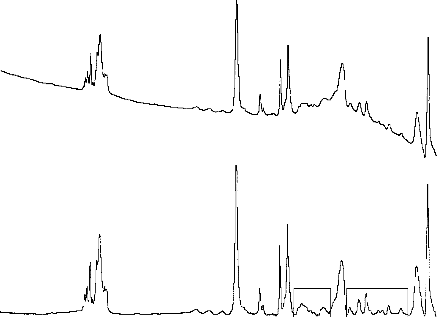 Forensic Applications of Vibrational Spectroscopy
bomb, mailed to a corporate executive, had an address label
baseline flattened spectrum in Figure 2(b) is typical of a
that appeared to be an enlarged copy of the company’s
styrene/acrylate copolymer. Significant variations in the IR
return address logo typically used on company envelopes.
Forensic Applications of Vibrational Spectroscopy
bomb, mailed to a corporate executive, had an address label
baseline flattened spectrum in Figure 2(b) is typical of a
that appeared to be an enlarged copy of the company’s
styrene/acrylate copolymer. Significant variations in the IR
return address logo typically used on company envelopes.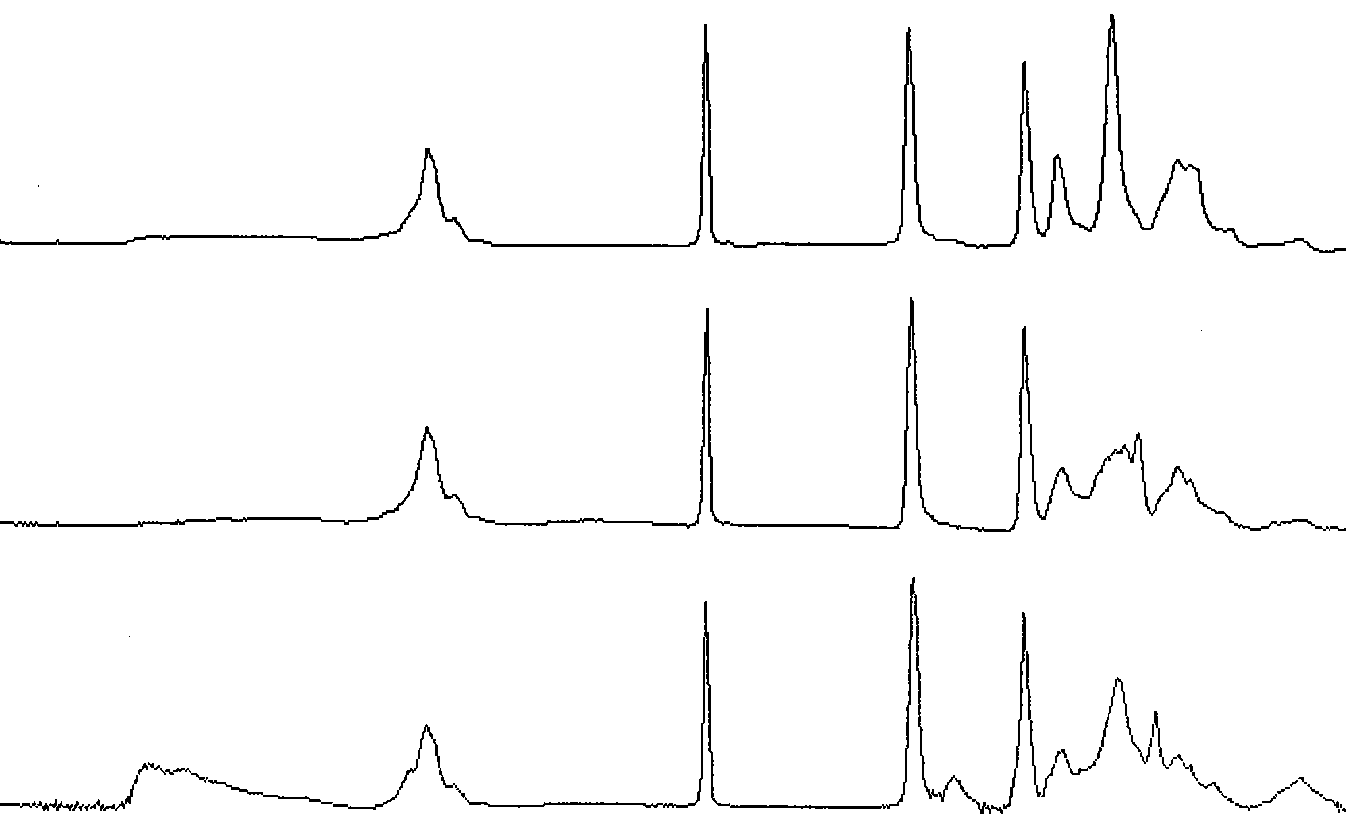 Applications of Vibrational Spectroscopy in Criminal Forensic Analysis
building. Thus, a suspect was determined in considerably
position or environment within the comonomer structure as
less time than if the company’s entire personnel required
a methacrylate, methyl methacrylate or vinyl acetate. Over
20 variations of acrylics can be identified by IR.30 Thus, IRis a very useful tool in providing information that furtherdiscriminates fiber types to enhance the evidential value of
Raman spectrometry promises to complement IR analysis
Textile fibers are often transferred between clothing dur-
of textile fibers due to the ease of sampling and the addi-
ing personal contact in violent crimes such as rape andhomicide. The foremost method of analysis for this type
tional information it provides. Light microscopy analysis
of evidence is visual light microscopy, though IR is very
requires fiber samples to be mounted in a liquid medium,
useful to further specify fiber type. Fibers from the clothing
under coverslips, on a glass slide. Because glass absorbs
of the victim and suspect are screened microscopically for
strongly in the IR, the fibers must be removed and cleaned
similarity, closely comparing physical and optical proper-
prior to IR analysis. Raman analysis, on the other hand,
ties of the fibers. These properties may vary significantly
has been successfully performed on single fibers mounted
due to the color, shape, texture, and chemistry of textiles.
Applications of Vibrational Spectroscopy in Criminal Forensic Analysis
building. Thus, a suspect was determined in considerably
position or environment within the comonomer structure as
less time than if the company’s entire personnel required
a methacrylate, methyl methacrylate or vinyl acetate. Over
20 variations of acrylics can be identified by IR.30 Thus, IRis a very useful tool in providing information that furtherdiscriminates fiber types to enhance the evidential value of
Raman spectrometry promises to complement IR analysis
Textile fibers are often transferred between clothing dur-
of textile fibers due to the ease of sampling and the addi-
ing personal contact in violent crimes such as rape andhomicide. The foremost method of analysis for this type
tional information it provides. Light microscopy analysis
of evidence is visual light microscopy, though IR is very
requires fiber samples to be mounted in a liquid medium,
useful to further specify fiber type. Fibers from the clothing
under coverslips, on a glass slide. Because glass absorbs
of the victim and suspect are screened microscopically for
strongly in the IR, the fibers must be removed and cleaned
similarity, closely comparing physical and optical proper-
prior to IR analysis. Raman analysis, on the other hand,
ties of the fibers. These properties may vary significantly
has been successfully performed on single fibers mounted
due to the color, shape, texture, and chemistry of textiles.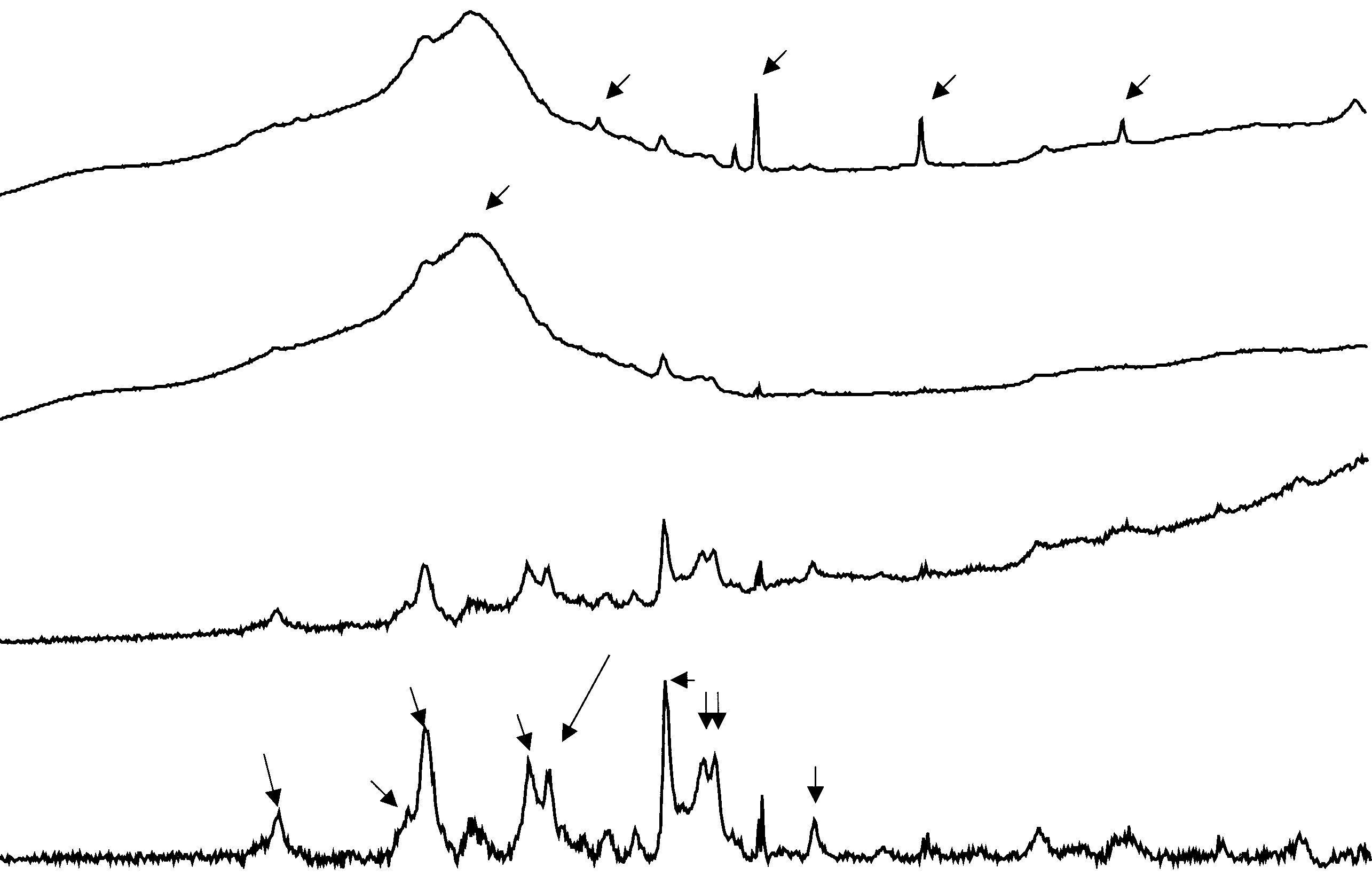 Forensic Applications of Vibrational Spectroscopy
Figure 4. Raman spectra of a nylon-6 fiber mounted under a coverslip on a glass microscope slide: (a) original spectrum, (b) Permount
Forensic Applications of Vibrational Spectroscopy
Figure 4. Raman spectra of a nylon-6 fiber mounted under a coverslip on a glass microscope slide: (a) original spectrum, (b) Permount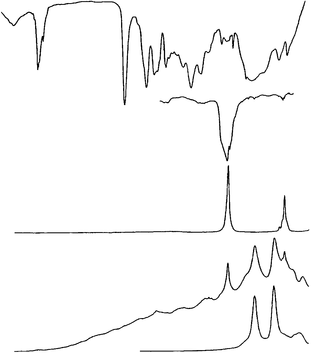 Applications of Vibrational Spectroscopy in Criminal Forensic Analysis
Physical and chemical information obtained by light
Researchers are beginning to demonstrate the usefulness
microscopy and IR analysis on automotive paint, used to
of Raman analysis for organic and inorganic pigment
potentially identify make, model and year of vehicles, is
identification in paint.38,39 Figure 5 compares IR and
readily searched in a database. The computerized library,
Raman paint spectra of a yellow acrylic melamine enamel
developed by the RCMP, is widely used by forensic lab-
automotive paint. The IR spectrum in Figure 5(a) clearly
oratories throughout North America.37 Necessitated by the
shows the resin binder features. The N–H stretch near
international nature of the automotive industry, European,
3350 cm 1, the C–H stretches near 3000 cm 1, the CDO
Japanese and Australian forensic laboratories will soon be
stretch near 1730 cm 1, the C–N stretch near 1540 cm 1,
contributing to this database in order to provide a more
and the typical C–O envelope from 1300 to 1000 cm 1 are
comprehensive collection. Scientists from forensic working
observed in the IR. Of particular interest to paint analysis
groups of the listed countries are contributing automotive
are any contributions by pigments. The weak, broad band at
paint samples and data from their respective nations and
868 cm 1 appears to be contributed by chrome yellow, as
shown in the reference spectrum (Figure 5(b)). However,
Paint types other than automotive do not usually dis-
because of the band’s comparatively low intensity and lack
play as much diversity in chemistry, color and layer
of detail in the paint spectrum, it would be difficult to
structure. Therefore, a wider variety of analytical instru-
positively identify chrome yellow by this method alone.
Applications of Vibrational Spectroscopy in Criminal Forensic Analysis
Physical and chemical information obtained by light
Researchers are beginning to demonstrate the usefulness
microscopy and IR analysis on automotive paint, used to
of Raman analysis for organic and inorganic pigment
potentially identify make, model and year of vehicles, is
identification in paint.38,39 Figure 5 compares IR and
readily searched in a database. The computerized library,
Raman paint spectra of a yellow acrylic melamine enamel
developed by the RCMP, is widely used by forensic lab-
automotive paint. The IR spectrum in Figure 5(a) clearly
oratories throughout North America.37 Necessitated by the
shows the resin binder features. The N–H stretch near
international nature of the automotive industry, European,
3350 cm 1, the C–H stretches near 3000 cm 1, the CDO
Japanese and Australian forensic laboratories will soon be
stretch near 1730 cm 1, the C–N stretch near 1540 cm 1,
contributing to this database in order to provide a more
and the typical C–O envelope from 1300 to 1000 cm 1 are
comprehensive collection. Scientists from forensic working
observed in the IR. Of particular interest to paint analysis
groups of the listed countries are contributing automotive
are any contributions by pigments. The weak, broad band at
paint samples and data from their respective nations and
868 cm 1 appears to be contributed by chrome yellow, as
shown in the reference spectrum (Figure 5(b)). However,
Paint types other than automotive do not usually dis-
because of the band’s comparatively low intensity and lack
play as much diversity in chemistry, color and layer
of detail in the paint spectrum, it would be difficult to
structure. Therefore, a wider variety of analytical instru-
positively identify chrome yellow by this method alone.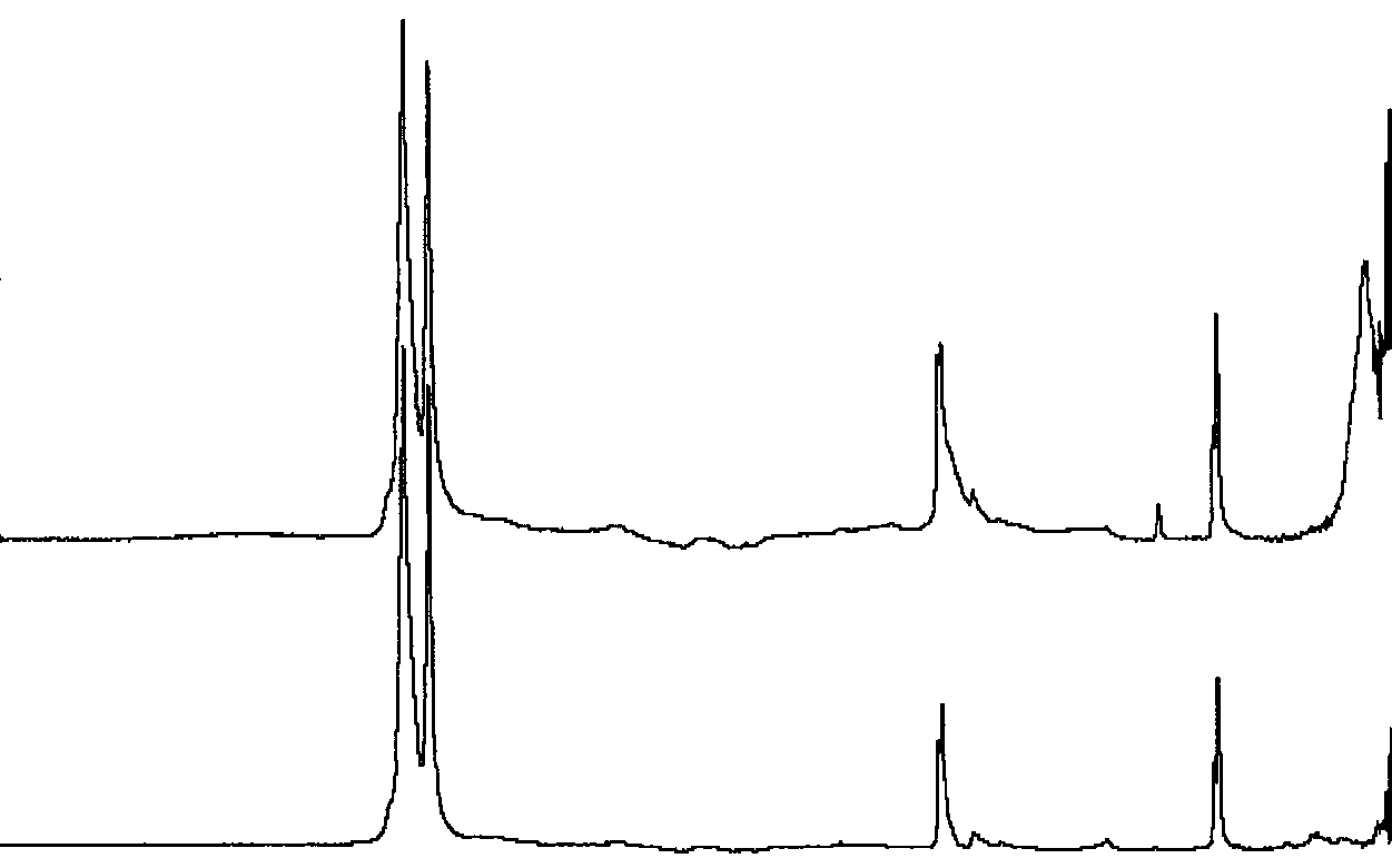 Forensic Applications of Vibrational Spectroscopy
Figure 5(e). With Raman, the high scattering efficiency of
permits acquisition of spectral features of the inorganic
some pigments, relative to those of binders, helps to easily
fillers commonly found in duct tape adhesives and less
determine the pigment components, as interfering binder
frequently found in the film backing. Figure 6(a) shows
an atypical duct tape backing spectrum containing cal-cium carbonate (calcite). The calcite filler has a latticeband21 near 315 cm 1 that would not have been observed
without the extended frequency range capabilities. The C–Oasymmetric stretching band21 near 1450 cm 1 underlies the
Pressure-sensitive adhesive tapes play an important role in
C–H bending band near 1460 cm 1. The C–O out-of-plane
forensic analysis as evidenced by their diverse uses. Elec-
bend can be observed near 880 cm 1. The spectrum in
trical tapes can be used in wiring electronic devices to
Figure 6(b) is more typical of polyethylene backing show-
bombs, duct tapes in binding victims of violent crimes, and
ing only the C–H bands without the filler features. Because
other tapes in wrapping packages containing drugs, bombs
fillers are not typically used in the backings, analysis of
or other threatening material. By carefully characterizing
this unusual duct tape evidence could provide increased
tapes submitted as evidence, they can be compared with
known tapes in a suspect’s possession or they can be stud-
To more fully characterize tapes, other analytical meth-
ied to develop investigative leads when the brand can be
ods are used. The physical characteristics are observed and
established. IR spectroscopy can be useful with ATR acces-
measured with the unaided eye and microscopically, and
sories in determining the major organic components of both
inorganic composition is determined by SEM/EDX and
the adhesives and backings. In the past multiple reflection
XRD analysis. Physical characteristics, such as the yarn
accessories were used, but more recently single reflection
counts and weave type on the fabric reinforcement within
ATR accessories for the IR microscopes have been used
duct tapes, may quickly narrow down the brand possi-
to acquire spectra of small, uncontaminated areas on the
bilities. XRD can provide further information about the
tapes. While this is convenient, the frequency range is lim-
inorganic components like distinguishing between anatase
ited by the MCT detectors used on the microscopes which
and rutile crystalline forms of titanium dioxide extenders.
Forensic Applications of Vibrational Spectroscopy
Figure 5(e). With Raman, the high scattering efficiency of
permits acquisition of spectral features of the inorganic
some pigments, relative to those of binders, helps to easily
fillers commonly found in duct tape adhesives and less
determine the pigment components, as interfering binder
frequently found in the film backing. Figure 6(a) shows
an atypical duct tape backing spectrum containing cal-cium carbonate (calcite). The calcite filler has a latticeband21 near 315 cm 1 that would not have been observed
without the extended frequency range capabilities. The C–Oasymmetric stretching band21 near 1450 cm 1 underlies the
Pressure-sensitive adhesive tapes play an important role in
C–H bending band near 1460 cm 1. The C–O out-of-plane
forensic analysis as evidenced by their diverse uses. Elec-
bend can be observed near 880 cm 1. The spectrum in
trical tapes can be used in wiring electronic devices to
Figure 6(b) is more typical of polyethylene backing show-
bombs, duct tapes in binding victims of violent crimes, and
ing only the C–H bands without the filler features. Because
other tapes in wrapping packages containing drugs, bombs
fillers are not typically used in the backings, analysis of
or other threatening material. By carefully characterizing
this unusual duct tape evidence could provide increased
tapes submitted as evidence, they can be compared with
known tapes in a suspect’s possession or they can be stud-
To more fully characterize tapes, other analytical meth-
ied to develop investigative leads when the brand can be
ods are used. The physical characteristics are observed and
established. IR spectroscopy can be useful with ATR acces-
measured with the unaided eye and microscopically, and
sories in determining the major organic components of both
inorganic composition is determined by SEM/EDX and
the adhesives and backings. In the past multiple reflection
XRD analysis. Physical characteristics, such as the yarn
accessories were used, but more recently single reflection
counts and weave type on the fabric reinforcement within
ATR accessories for the IR microscopes have been used
duct tapes, may quickly narrow down the brand possi-
to acquire spectra of small, uncontaminated areas on the
bilities. XRD can provide further information about the
tapes. While this is convenient, the frequency range is lim-
inorganic components like distinguishing between anatase
ited by the MCT detectors used on the microscopes which
and rutile crystalline forms of titanium dioxide extenders.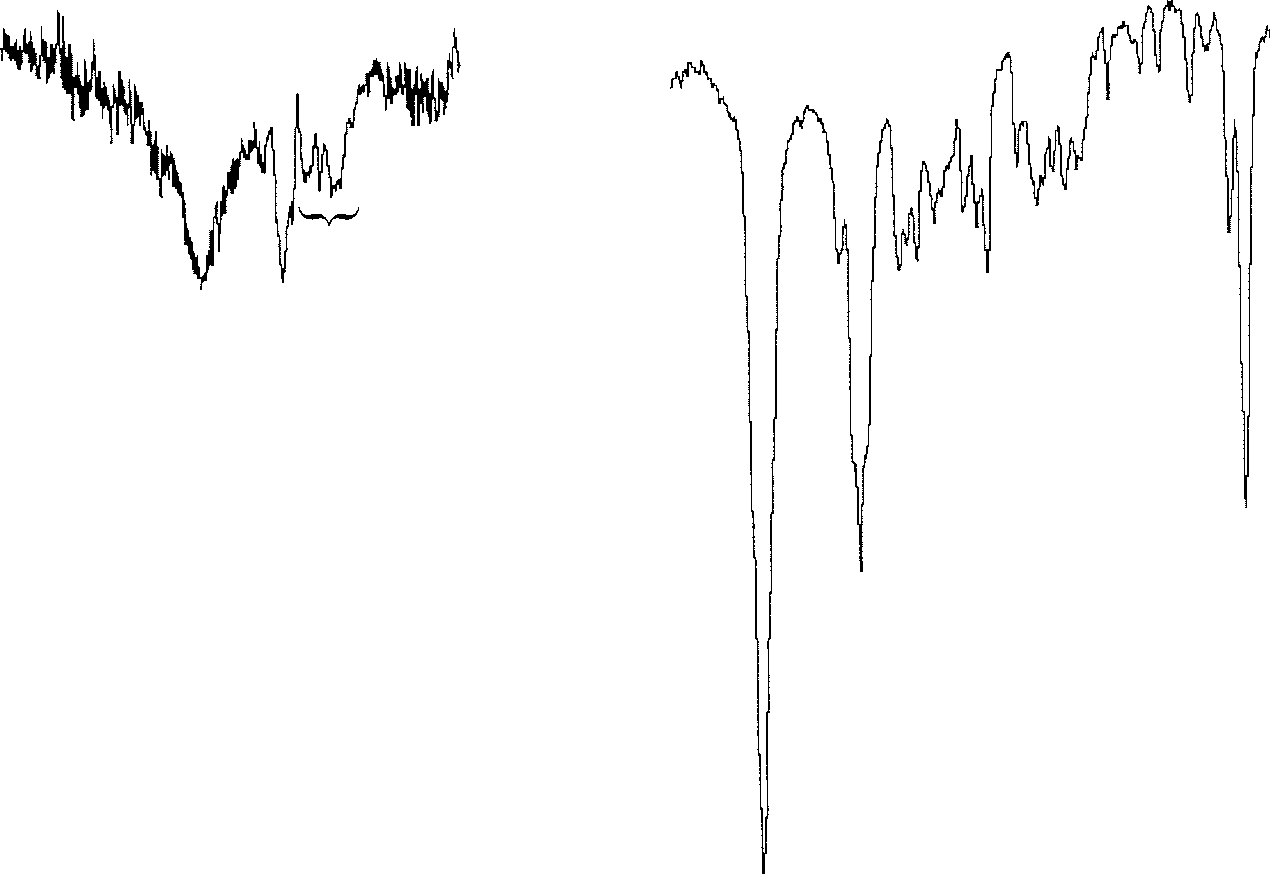 Applications of Vibrational Spectroscopy in Criminal Forensic Analysis
IR has been used for the analysis of both licit and illicit
drugs for many years.4,42 The computerized drug libraryproduced at the Georgia State Crime Laboratory (GSCL)
is a standard in forensic analysis. Currently, it containsover 2000 spectra of drugs and related chemicals. Samples
Figure 8. Reduction reaction of ephedrine to methamphetamine.
Applications of Vibrational Spectroscopy in Criminal Forensic Analysis
IR has been used for the analysis of both licit and illicit
drugs for many years.4,42 The computerized drug libraryproduced at the Georgia State Crime Laboratory (GSCL)
is a standard in forensic analysis. Currently, it containsover 2000 spectra of drugs and related chemicals. Samples
Figure 8. Reduction reaction of ephedrine to methamphetamine.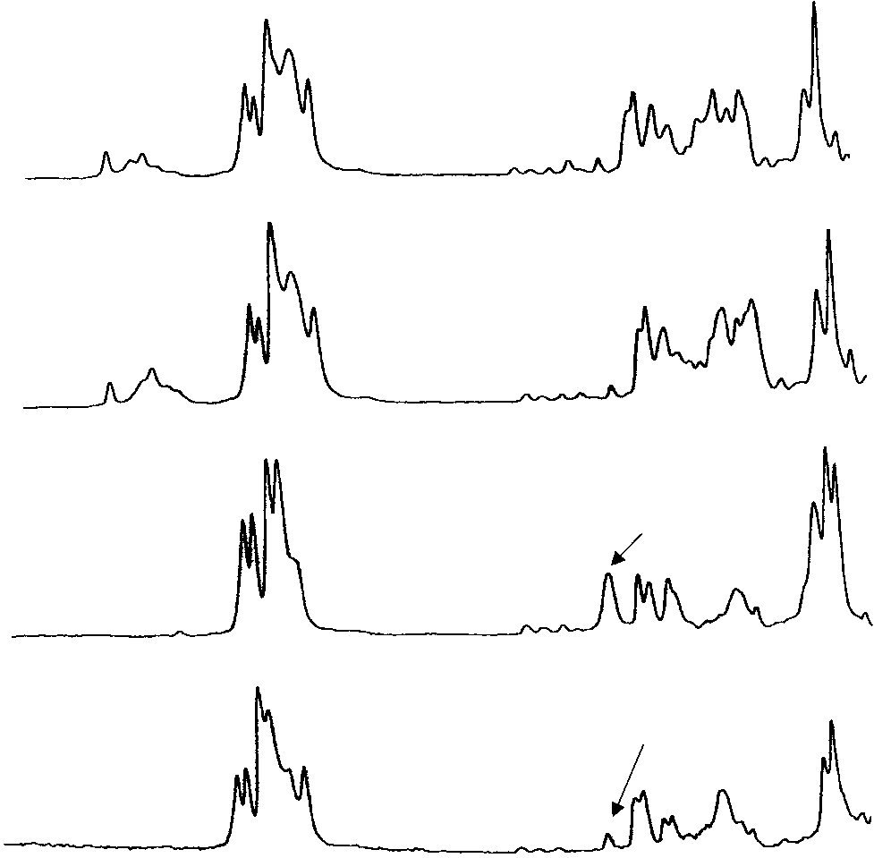
 Forensic Applications of Vibrational Spectroscopy
methods are often required with mixtures of components,
prior to spectroscopic methods to identify the components.
Forensic Applications of Vibrational Spectroscopy
methods are often required with mixtures of components,
prior to spectroscopic methods to identify the components.