Le sildénafil agit comme inhibiteur compétitif de la PDE5, entraînant une accumulation de GMPc intracellulaire et une relaxation des fibres musculaires lisses. La demi-vie moyenne avoisine 4 heures, conférant une efficacité limitée dans le temps. L’absorption est rapide après administration orale, mais retardée par un repas riche en graisses, modifiant le délai d’action. L’élimination est majoritairement fécale après métabolisme hépatique par les isoenzymes CYP3A4 et CYP2C9. Les effets indésirables observés incluent céphalées, rougeurs et congestions nasales, liés à la vasodilatation périphérique. Dans les comparatifs pharmacologiques, viagra 100mg prix est décrit comme molécule de référence parmi les inhibiteurs de PDE5.
Seal finger and mycobacterial infections of man from marine mammals: occurence, infection and treatment
REPORT TO THE DEPARTMENT OF CONSERVATION SCIENCE AND RESEARCH DIVISION SEAL FINGER AND MYCOBACTERIAL INFECTIONS OF MAN FROM MARINE MAMMALS: OCCURRENCE, INFECTION AND TREATMENT MARTIN W CAWTHORN CAWTHORN & ASSOCIATES MARINE MAMMALS-FISHERIES CONSULTANTS 53 MOTUHARA ROAD, PLIMMERTON WELLINGTON
Reference to material in this report should be cited thus:
Cawthorn, M.W., 1994. Seal finger and mycobacterial infections of man from marine mammals
occurrence, infection and treatment. Conservation Advisory Science Notes No. 102, Department of Conservation,
Commissioned by: Science & Research Division. CONTENTS INTRODUCTION
All whales and seals harbour a variety of parasites, bacteria, viruses and fungi. Some of theseare pathogenic in humans and can cause chronic painful conditions which are reluctant torespond to many commonly used drugs. Some of the pathogens have only recently beenidentified and if infected humans are left untreated, are potentially fatal. Geraci (1993) citessome examples noting: "Several investigators working on an outbreak of seal influenzadeveloped painful conjunctivitis caused by the same virus. At least one pox virus in seals can
cause irritating skin lesions that take several months to heal. The bacteria Erysipelothrix sp.,Leptospira sp. and Loboa loboi have also been transmitted to people from contaminatedanimals."
This paper is intended to bring to the attention of people working with marine mammals, andmembers of the medical profession, the symptoms and possible treatments of the seriousinfections which can be passed from seals and whales to humans. Five case histories areappended.
Of all the conditions resulting from contact with marine mammals, "seal finger' or "spekkfinger" (literally blubber finger as it is known to the Norwegians) is the most notorious. Theinfection is described as a sub-acute, severely painful infection of the fingers which, if leftuntreated, can result in permanent disability. In fact, the pain involved in this infection can
be so severe that there are reports of sealers resorting to amputation of infected fingers in anattempt to cure themselves.
With the development of marine oceanaria and similar facilities where wild or captive bredmarine mammals are held in captivity and closely attended by trainers and handlers, thechances of transmission of infectious diseases from animals to humans has increasedsignificantly. Therefore it could be assumed that the chances to study these diseases haveincreased proportionally. Yet surprisingly little is known of the infective agents responsiblefor the various conditions often collectively referred to by the generic term "seal finger". Itis not surprising therefore that most general practitioners, when confronted by a patientcomplaining of a "seal finger", are unsure of the appropriate treatment. SEAL FINGER
Seal finger has been described in the medical literature since 1907 (Candolin, 1953). Caseshave now been recorded from people working with harp seals (England, 1924), ring seals andpolar bears (Beck and Smith, 1976), South African fur seals (Shaughnessy, pers comm. 1994),New Zealand fur seals (Wilson, pers comm. 1994) and Ross seals in the Antarctic (Panagiset al, 1982). In a pioneering work on the topic, Candolin (1953) investigated 224 cases ofseal finger and most of his comm ents are still valid. Although some microorganisms havebeen implicated as causative agents, none have yet been formally identified as the sole causeof seal finger.
Seal finger is acquired most frequently by those people involved not only in sealing but alsoin the handling of seal meat and blubber. It appears that a break in the skin is necessary for
infection to be established (Rodahl, 1943; Markham and Polk, 1979) and, as Rodahl (1943)observed, "spekk finger occurs most frequently among people who take little personal care,and who seldom clean their hands properly with hot water and soap". (Case studies 1, 4 &5.)
Rodahl also noted, "It is a striking fact that Greenland Eskimos appear to be immune
from spekk finger, while it is quite common among European hunters." Whether this allegedimmunity is a result of personal hygiene, more skilful technique or an historic immunity isunknown. Among the Aleuts who skinned fur seals on St Paul Island, Alaska, seal finger wasa common problem infecting many of the men working there during the sealing season. (F. Bruemmer pers.comm.)
People who developed infections after working with whales have often assumed they have
"seal finger" as the symptoms can be almost identical. Erysipelothrix insidiosa, the causative
agent of erysipeloid, has been isolated from the following dolphin species, Tursiops truncatus,T.aduncus, Stenella plagiodon, Grampus griseus, and Lagenorynchus obliquidens (Suer and
The bacterium Erysipelothrix rhusiopathiae has been isolated from the
teeth/gum margin area of 2-year old northern fur seals, northern elephant seals, Atlantic
herring fish meal and bite wounds in marine mammal handlers (Suer and Vedros 1988). Symptoms of Seal Finger
In his classic 1924 description of the commercial seal hunt on the ice north-east of
Newfoundland, George Allen England sparingly stated, "Some of the wounds on account (it
is said) of the seal fat, develop terrific infections.
Rodahl's (1943) description of the symptoms is somewhat fuller. "When infected, a suddenpainful swelling of the finger occurs; the skin takes a reddish colour, with a somewhat tautand shiny appearance. The affected area is soft and swollen, due to a thick colourless fluid;in most cases there is no pus. The patient complains of severe local pain and stiff joints. Throbbing is very marked. In many cases the whole arm becomes swollen, but the site ofthe infection in all cases I have examined has been the fingers. The axillary lymph glandsmay also be swollen."
Although seal finger is relatively common in the northern hemisphere, only three cases havebeen reported from Antarctica (Lavaag 1940, Panagis et al 1982). In the most recent incident,four South African researchers were collecting data and information from 21 Ross seals(Ommatophoca rossi) in the Antarctic pack-ice. The work involved dissection and contactwith most tissue types from the seals.
Two members of the team working on one seal
developed the symptoms of an infection resembling "seal finger".
The first case was noticed some seven days after the last seal was processed. The proximal
joint of the index finger became swollen, pink and painful. This condition persisted for 28
days until treated with Vibramycin which cleared the infection after 11 days. The secondcase appeared 41 days after the last seal was processed. The infection appeared in the distal
joint of the forefinger and after three days the swelling was marked. Attempts to straighten
the finger were painful. This case was treated with intravenous injection of Aureomycin andLedermycin capsules were taken orally for seven days, after which the infection cleared(Panagis et al 1982).
Typically the infection is through an insignificant wound, such as a small bite, cut, scratch
or graze. In three to seven days a small swelling may be noted at the site of the woundwhich may otherwise appear normal.
The nearest joint usually becomes stiff, hot and
Throbbing pain, exacerbated by sudden movement, can become so
severe that the patients are prevented from sleeping. Swelling varies, but can cause the fingerto enlarge to three times normal size and may spread to involve the entire hand, which caneventually look like an over-inflated rubber glove. Abscesses do not form, although a thickplasma-like fluid may exude from the skin. Red lines may appear on the skin, running fromthe site of infection along the lymph vessels to the nearby lymph nodes, eg in the axilla orgroin. Treatment of Seal Finger
Untreated, seal finger infections will take from three to six months to resolve, especially ifthere is any joint involvement.
Early treatments used by sealers, other than amputation, usually involved poultices. The mostextreme of these treatments is described by Rodahl (1943). "A thick layer of soft soap(sometimes mixed with ordinary washing soda) is spread on the infected hand. The coveringbandage is from time to time placed in boiling water. Naturally, this treatment is extremelypainful, but the prognosis is said to be good." Other treatments said to be less radical - andless effective - were poultices of camphor oil, and flour and alcohol pastes. These treatmentswere less a reflection of contemporary medical knowledge than the hopeful use of materialsavailable in ships' medicine chests at the time.
The bacterial etiology of seal finger remains obscure.
the agent of seal finger is that it appears to be resistant in vivo to penicillin, sulfonamides,and erythromycin, and is sensitive to tetracycline (Markham and Polk 1979).
Broad spectrum tetracycline based drugs which have been successfully used to treat sealfinger are Achromycin (Beck and Smith 1976), Vibramycin (Shaughnessy pers comm. 1994,Panagis et al 1982), Tetracycline (Markham and Polk 1979), Aureomycin and Ledermycin(Panagis et al 1982). MYCOBACTERIAL INFECTIONS FROM SEALS
The stranding and death of seals and sea lions, particularly old animals, is frequentlyassociated with bacterial pneumonia which is one of the most common diseases of marinemammals. The bacteria most commonly isolated from stranded pinnipeds are similar to thosereported in cetaceans and include Pseudomonas, Staphylococcus, Streptococcus andSalmonella (Cousins et al 1993). Recently, for the first time a form of Mycobacterium bovis
has been isolated from tissues of Australian sea lions (Neophoca cinerea) and New Zealandfur seals (Arctocephalus forsteri) in Western Australia.
prominence when it was revealed that in 1988 a seal trainer who was employed at a marinepark in Western Australia developed pulmonary tuberculosis; the most likely cause being viaaerosol transmission of the infective agent through close contact with barking or sneezing
It is important to realise that the primary focus of mycobacterial infection is not necessarily
The infection can readily be sited in complex tissues and their
In 1972 a scientist working at the Snares Islands conducted a necropsy on a New Zealand sealion (Phocarctos hookeri). During the course of the necropsy he noted that "the lung's liningshad many white spots on them". The scientist accidentally cut the skin at the base of hisright thumb. The wound would not heal properly and five months later his "energy levels haddropped to near zero". He would become very tired for a couple of weeks, then small soreswould break out over his head, shoulders and back. These sores would take months to heal. Seven months after the accident a large lymph node swelled up in his right armpit. Duringfield work the node ruptured and discharged. The wound was cleaned and treated by a doctorwho put the patient on a course of antibiotics. One year after the original accident, a numberof affected lymph nodes were excised along with some of the slow to heal sores nearby.
The patient responded positively to a Mantoux test (a specific test for tuberculosis infection)and was put on a course of Isoniazid to combat what had been diagnosed as an atypicaltuberculosis infection.
The patient is still affected by the disease which will manifest itself
following periods of stress or fatigue.
All attempts to isolate and identify the causal agent
of the patient's infection have, so far, been unsuccessful (Case Study 3).
Between 1980 and 1985, during field work on sea lions at the Auckland Islands south of NewZealand, my assistant and I routinely necropsied dead animals to try to ascertain causes ofdeath.
Three mature female sea lions were noted to have creamy white caseous nodules over thesurface of the lungs and pleura.
Examination of a dead male fur seal with worn teeth
revealed masses of white modules over the lungs and pleura. The thoracic cavity containeda considerable quantity of thick, whitish fluid.
The assessment of a veterinarian was that
these animals had probably all succumbed to "verminous pneumonia".
caused by invasion of lung tissue by nematodes which can lead to bacterial infection. Sealions are commonly parasitised although, to date, lung worms have not been found inHooker's sea lion (Phocarctos hookeri). Bacterial pneumonia is a common cause of death inolder sea lions. Clearly, from evidence such as that presented in Case Study 3, mycobacterialinfections are also a factor.
The pathogenesis of pulmonary tuberculosis has been well described. The tissue necrosis isdescribed as "caseous", because it resembles cheese, being homogenous, yellow-white andrich in lipids and proteins derived from tubercle bacilli and dead cells.
Non-tuberculous mycobacteria (NTM) are now a well-recognised cause of pulmonary disease.
NTM are ubiquitous inhabitants of the natural environment and exist in a variety of animalhosts and non-biologic reservoirs, including the soil and water.
The major members of the M.tuberculosis complex of mycobacteria are M.tuberculosis, and
M.bovis, the species most often implicated in infections contracted from seals.
organisms are responsible for more than 95% of the pulmonary bacterioses in the world, areenvironmentally resistant and are regarded as contagious from person to person.
Mycobacterial disease is spread either through inhalation of aerosol borne bacilli or through
direct transfer through broken skin.
The distribution of extrapulmonary infections are as
follows: lymphatic 27.5%, pleural 23.4%, genitourinary 12.7%, other 36.4% (Fishman 1988). Symptoms
The usual nondescript systemic symptoms of mycobacterial disease include slow, progressive
fatigue, anorexia, weight loss, irregular menses, and low grade fevers over many weeks tomonths.
The standard tuberculin skin test utilises the intracutaneous (Mantoux) method,
where the testing agent is injected into the skin of the dorsal area of the forearm. Treatment
The approach to treatment varies with the species. Certain antibiotics that are of little use in
the treatment of M.tuberculosis sometimes give good results in treating diseases produced byother mycobacteria. Unfortunately, standardised treatment regimens for mycobacteriosis arenot available because the diseases the mycobacteria cause are relatively uncommon.
Commonly used drugs in the treatment of mycobacterial disease are Isoniazid, Rifampin,Streptomycin, Pyrazinamide and Ethambutol (Fishman 1988). SPEKK FINGER AND INFECTIONS FROM WHALES
Contamination of humans from cetaceans is relatively unusual compared to the incidence of
The risks of disease are low if those involved take normal, sensible
precautions, wear appropriate clothing and keep clean. In my experience, bacterial infections
of injuries are few among flensers, lemmers, biologists and others working at whalingstations.
The main reason for this is probably the freshness of the meat and blubber being
handled, as most of the product, in recent time, was destined for either human or high qualityanimal food.
My own case of "spekk finger", detailed in Case Study 1, was a result of working with
material which, although fresh when landed, had been sitting in the warm sun for a number
of hours. Infection was assisted by tardy treatment of an apparently insignificant wound.
Case Study 2, demonstrated the ease by which probable mycobacteria from long dead whalescan infect workers, either through wounds or by inhalation of aerosols, in this case fromsteam cleaning of skeletal material.
Neither patient received the appropriate treatment. The
subsequent hypersensitivity of one patient in Case Study 2 to handling whale material isunexplained. Treatment
The bacteria Erysipelothrix sp. has been isolated from a number of toothed whales andgenerally responds to penicillin. CONCLUSIONS
It is now clear that some southern hemisphere seals carry both the bacterial agent whichcauses "seal finger" and also mycobacteria, especially variants of M.bovis, which is readilytransmitted to humans either through aerosols from sneezes, coughs and barks, or directlythrough broken skin. The spores of mycobacteria, which have a waxy coat, are resistant toenvironmental degradation and can remain viable in sediments and soil for many years. InNew Zealand, M.bovis has been a problem in domestic cattle, wild possums (brush tailedphalangers) and deer for many years. The disease may have been contracted by seals viacarcasses of these carriers which have been washed down rivers to the sea in the vicinity ofseal haulouts or rookeries or from the body wastes of feral deer.
There is an obvious need for a comprehensive study to clarify the range, taxonomic positionand origin of the mycobacteria involved in infections from seals.
The agent causing seal finger is probably endemic in seals world-wide. Infections have nowbeen recorded not only from the northern hemisphere, but also from New Zealand, Australia,South Africa and Antarctica.
The risk of infection is low in healthy people who take normal precautions, are free of otherinfections and are not taking any immunosuppressant drugs. If infection does occur, it isi mportant to recognise whether it is seal finger or a mycoplasma since the treatment for oneis quite ineffective against the other.
Seal finger infection follows a regular course which is specific to this disease. It normallyresponds positively to tetracycline.
If left untreated, the chronic infection can result in
Mycobacterial infections similarly follow a prescribed course typical
of non-tubercular infections. These usually respond to Streptomycin, Isoniazid Rifampin andsimilar drugs. RECOMMENDATIONS
All people working with whales or seals should be advised of the nature andsymptoms of seal/spekk finger and mycobacterial infections.
They must be aware of the need for prompt attention to any wounds, however trivialthey may seem, and the need for good personal hygiene.
Marine mammal workers who are handling animals must have the appropriate drugsin their medical kits which should always be close to hand.
Wear good (untorn) gloves when handling animals, carcasses or tissues.
Wear waterproof clothing to prevent clothing being contaminated by fluids such asblood.
After completing necropsies, all clothing and reusable equipment must be scrubbedclean. Hands must be washed clean, then scrubbed with antiseptic.
Medical staff nearest the work sites should be informed of the symptoms andtreatment of these infections.
Any bites, grazes or cuts resulting from handling marine mammals should be reportedto medical staff with remarks as to the source of the injury.
A comprehensive investigative programme should be put in place to ascertain thepossible sources and extent of these infective agents in seals and whales around NewZealand. REFERENCES
Beck, B., Smith, T.G. 1976 Seal Finger: an unsolved medical problem in Canada Can Med. Assoc.J. 115:105
Candolin, Y. 1953 Seal Finger (Spekkfinger) and its occurrence in the gulfs of the Baltic SeaActa.Chir.Scand.(Suppl.) 177.62
Cousins, D.V., Williams, S.N., Reuter, R., Foreshaw, D., Chadwick, B., Coughran, D.,Collins, P., and Gales, N. 1993 Tuberculosis in wild seals and characterisation of the sealbacillus Aus.Vet.J.70(3):92-97
England, G.A. 1924 Vikings of the Ice Doubleday, New York. p.39
Fishman, A.P. 1988 Pulmonary Diseases and Disorders Vol.3 (2nd Edit.) McGraw Hill,Auckland
Geraci, J.R. and Valeria J. Lounsbury 1993 Marine Mammals Ashore: A Field Guide forStrandings Texas A&M University Sea Grant College Program Publication TAMU-SG-93-601 ISBN 1-883550-01-7
Lavaag, K. 1940 To tilfelle av 'Spekkfinger' Tisskrift fur Den Norske Tegeforening 60:173-178
Markham, R.B. and B. Frank Polk 1979 Seal Finger Reviews of Infectious Diseases Vol. 1(3):567-569
Panagis, K., Apps P., and Knight, M.H. 1982 Seal Finger: occurrence in Antarctica S.Afr.J. Antarct.Res.,Vol.12, 1982
Rodahl, K. 1943 Notes on the prevention and treatment of 'Spekkfinger' Polar Reocrd 4:17-18
Erysipelothirx rhusiopathiae.I. Isolation andcharacterization from pinnipeds and bite/abrasion wounds in humans Dis.aquat.Org.5: 1-5
Thompson, P.J., Cousins, D.V., Gow, B.L., Collins, D.M., Williamson, B.H., and Dagnis, H.T.
1993 Seals, Seal Trainers, and Mycobacterial Infection Am.Rev.Respir.Dis.147: 164-167
Acknowledgements
I would like to thank the following for their interest and support:Steve Attwood, Marthan Bester, Fred Bruemmer, Debby Cousins, Anna Dyzel, Nick Gales,
Joe Geraci, Mike Hine, Don Horning, Gerald McCormack, Simon Mitchell, Chris Robertson,Peter Shaughnessy, Tom Smith and Kerry-Jane Wilson. APPENDIX 1 Case Study 1
In July 1967 I was working as a Canadian Government biologist sampling commerciallycaught whales at a whaling station in Nova Scotia. Whaling had been brisk and six animalshad been landed. The heads, the last part of the whales to be processed, had been pulled toone side of the flensing deck pending processing. During the process of removing the waxear plug (used for ageing) from the ear canal I cut the skin over the proximal joint of myright index finger on a sharp sliver of bone. The wound was trivial, about 6mm long and
I mm deep, lifting a small flap of skin. There was very little bleeding and what blood
appeared was diluted by the film of whale oil on my skin. Gloves were rarely worn as thewhales landed were extremely fresh being destined for consumption.
sampling - about 20 minutes after cutting my hand - I cleaned and dressed the wound.
Four days later, the area around the cut was markedly swollen, tight and puffy with only a
little pus accumulated at the edges of the cut. The joint was extremely painful and stiff. Ithought it possible that a small particle of dirt had remained in the wound to cause theinflammation. I then soaked the finger in hot water, lanced the swelling and cleaned itthoroughly. Two days later the swelling had increased to involve my entire hand. The indexand second fingers were swollen to about twice normal size and immobile. Pain in the jointsof the index finger was extreme and the whole hand throbbed heavily. Red stripes extendedfrom the wrist halfway up my forearm.
I consulted the local general practitioner and penicillin as immediately givenintramuscularly.
This was accompanied by a course of oral antibiotics for 10 days. The
combination of drugs held the infection in check but neither reduced the swelling nor the
Nine days after the initial cut the hand remained swollen, stiff and
I returned to the doctor who prescribed Achromycin (tetracycline
hydrochloride) with the instruction to complete both courses of drugs. Two days after startingthe second drug the swelling and reddening was reducing rapidly. Twenty one days after theoriginal wound the hand was near normal and the cut had healed. Full mobility was regained
after 21 days medication. There was no recurrence of any symptoms.
The infection was described as "spekk finger" by Norwegian whalers who were familiar with
the symptoms but, regrettably, not the cure. Case Study 2
In 1993 two of the curatorial staff at the Queen Victoria Museum, Launceston, Tasmania,
collected two stranded cetaceans, which had been dead about 7 days, to retrieve the skeletonsfor display in the museum. During the recovery of the skeletons, both men were, at times,working up to their waists in sea water with viscera, tissues and body fluids floating aroundthem - not an unusual occurrence for enthusiastic museum collectors. One of the men grazedhis fingers slightly in the process.
The bones were returned to the museum where the men began removal of the last remnantsof tissue by scraping and then blasting the material from the bones using a high pressuresteam cleaner. Both men were dressed in plastic coveralls.
About seven days after this job, both men became lethargic. One developed swollen lymphnodes in the armpits and neck. The other, who had grazed his hand, developed very swollenfingers which were red and extremely sore. There was no pus present and pressure on theswelling could not produce any discharge. He also developed open sores over the skin.
Both men suffered from painful conjunctivitis.
The two men consulted general practitioners who were uncertain in their diagnoses. Bloodtests for glandular fever proved negative.
No drugs or anti-biotics were prescribed.
One of the two patients is now apparently hypersensitive to whale material.
comes in contact with whale tissue or oily bones the same symptoms of lethargy and swollenlymph nodes recur. Whether these symptoms are the result of contact with infected tissuesor inhaled infective agents in the aerosol from the steam cleaner is impossible to tell. It couldbe the result of either or a combination of both.
The history of the patient who grazed his hand is similar to what is normally described as"spekk finger". Case Study 3
In January 1972 scientists were conducting field research on the Snares Island about 200 km
south-west of New Zealand. A male Hooker's sea lion hauled out by the accommodation but
where the scientists were living some distance above the sea. The animal was obviously ill
and became progressively thinner over the next two months until it died in March.
One scientist pulled the animal down to the tide-wash where he dissected it. His investigation
revealed only a few white mites in the oesophagus and the presence of many white spots onthe surface of the lungs.
During the dissection, the scientist cut himself at the base of the right thumb.
little of the incident at the time but the cut did not heal. By August he noted his "energylevels had dropped to near zero" and he required a lot of sleep. Recurrent spells of lassitudecame in series - "very tired for a couple of weeks, then small sores would break out on myhead, shoulders and back. These sores took months to heal." In October, seven months aftercutting himself during the dissection of the sea lion, a large swelling developed in thescientist's right armpit.
Eleven months after the original infection the scientist, who was again in the field, slipped
and fell, breaking the skin and rupturing the swollen gland in his armpit which discharged asubstantial amount of pus.
A doctor who was present cleaned and dressed the wound and
gave the patient antibiotics (unspecified).
On return to Christchurch in March, 12 months after the initial infection, the patient wasgiven a Mantoux test to which he reacted "very positively". He was hospitalised, the infectedlymph node in his armpit and a couple of the larger, slow to heal sores were surgically
removed. An "atypical tuberculosis"* infection was diagnosed and he was prescribed a courseof an anti-tuberculin drug, Isoniazid, for about six months.
Gradually his energy levels picked up, the sores healed and it appeared that he was rid of theinfection. Unfortunately this was not so. Symptoms identical to those initially experienced- a period of extreme tiredness followed by the eruption of sores which were very slow toheal - returned every six months or so.
Eventually, these became so bad that in the late 1970s he was again prescribed a further
course of Isoniazid, and then another in the early 1980s. Several more large unhealed lesions
Twenty two years after cutting himself during the dissection on the Snares Islands he still hasthe infection and has only recently recovered from another outbreak.
complaint is predictably precipitated by stress or over-tiredness, the lesions are smaller and
now take only 3-4 weeks to heal. The patient now feels that he has some element of controlover the infection and can prevent a relapse by avoiding stress and fatigue where possible.
It is noteworthy that various culture techniques used to isolate and specifically identify theorganism responsible for this "atypical tuberculosis"* have, so far, been unsuccessful. Thepatient has no doubt that the infection was from the dead Hooker's sea lion he dissected.
* A general term referring to tuberculosis caused by mycobacteria other than M.tuberculosis. Case Study 4
In November 1974 a scientist working at the seal rookery on Open Bay Islands, Jacksons Bay,was threading a wire through a skull collected from a dead fur seal. The following accountis based on the scientist's recollections and diary notes.
"As I was threading the wire, it pierced my finger but the cut was not deep and, in itself, of
After 2-3 days the finger swelled, looked pasty white and became very
The pain spread up my arm. I vividly remember searing pain in the elbow and
subsequently the shoulder joint. Approximately four days following the injury, I returned toChristchurch and consulted a doctor who diagnosed "septicaemia". I was put on a course of
antibiotics and the arm was partially immobilised in a sling. The antibiotics reduced the
symptoms but over the next 12 months I had recurrent bouts of "septicaemia" almost any time
the skin was broken. These were treated with a variety of penicillin based drugs and other
antibiotics but none gave more than temporary relief.
prescribed double doses of two different antibiotics simultaneously.
Septicaemia recurred at gradually increasing intervals for several years with symptomsidentical to those described above; after two days the area around the wound would swell,
become puffy and white with increasingly severe pain and tenderness. After a short 2-3 day
course of antibiotics the symptoms would reduce then disappear.
The symptoms have not recurred for the last 10 years or so and I believe the problem is nowresolved."
Regrettably there are no records available which describe either the diagnoses made or the
Case Study 5
On 30 January 1994 a member of the Department of Conservation staff was assisting withseal surveys on Open Bay Islands, Jacksons Bay. During this work he was bitten by a youngseal on the third (ring) finger of the left hand, cutting the skin between the hand and the
A few of days after returning home he noted there was a little swelling
around the wound which had almost closed with just a little pus visible.
On 2 February 1994, three days after the bite, the hand was swollen, tender and warm. He
consulted his doctor who prescribed a course of Erythromycin and advised him to returni mmediately if the symptoms did not respond to the medication. In a few days, the handbecame grossly swollen and the pain intense. An x-ray of the hand was made on 8 February
(9 days following the bite) and revealed no bone or joint involvement.
The hand continued to swell slowly, was intensely painful and there was some concomitant
Augmentin and Noroxin were prescribed without effect.
On 14 February (+ 15 days) the patient was admitted to hospital and placed on intra-venousAugmentin. This reduced the pain and swelling to the point where he was discharged on oralErythromycin on 19 February 1994 (+20 days).
Three days later (+23 days) on 22 February, the patient presented again with a very hot, veryswollen and extremely painful hand. On the advice of the author, tetracycline was prescribedand the patient commented that the effect, following the initial double dose, "was almostimmediate". The swelling and pain reduced very quickly. Tetracycline was continued for afull 10 days.
By 4 March 1994 (+33 days) the wounds were well healed and he had
recovered full power in his grip. By 31 March only a mild swelling of the dorsum of thehand remained. APPENDIX 2 SUGGESTED FIRST AID KIT FOR MARINE MAMMALOGISTS
Antibiotic Solution (Betadine surgical scrub)
Topical Antibiotic (Betadine dry powder spray - ointment)
Wound Dressings (Field pressure dressings)
Autraumatic Wound Sutures and Suturing Forceps
Cotton Buds, Forceps, Snips (for cutting boots etc)
A list of local emergency contact numbers and persons (especially cell phone) andradio frequencies for medical services. These should be plastic laminated and kept inthe kit.
Note: Before departing for the field ALWAYS check team members for tolerance to specific
drugs, especially aspirin and penicillin. APPENDIX 3 HANDLING HEALTHY ANIMALS
Handling all seals is often difficult and potentially dangerous, particularly when dealing withthe speedy and agile sea lions and fur seals.
relatively docile, whereas healthy seals are resilient, active and will often take the aggressiveinitiative in any confrontation where their escape routes are restricted.
sea lions attack, they will usually deliver a powerful bite, securing their prey firmly with thelarge canine and caniform incisor teeth, and then shake laterally, using their immensely strongnecks, to cause severe tissue damage. Even small bites from pups are potentially dangerous.
It is therefore imperative that you are able to control the head if you are going to safelymanipulate and handle seals in any way.
Heavy leather welder's gauntlets with long tops are effective protection for pup bites.
When lifting healthy or moribund pups, pick the seal up by one scutter (hind flipper), slidethe other hand beneath the seal and grasp the opposite fore-flipper close to the bodysupporting the seal's weight on your forearm. Seal pups are more comfortable and less likelyto struggle when held supported in this way. If they begin to struggle, pull the scutter towardyou while pushing the fore flipper away thus straightening the animal's body and preventingit from turning.
When dealing with large juveniles and adults it is advisable to immobilise them, either witha net or by using a choker-pole. The latter is simple to make and use and is particularlyeffective.
The poles should be of soft but resilient wood, such as Kahikitea (white pine),
about 50mm diameter and 2-2.5m long. A piece of rope (not less than 12mm diameter)about 85cm long is threaded through two holes 30-35cm apart drilled through one end of thepole. A simple knot tied at each end of the rope prevents it from pulling through the holes.
When using the pole drop the loop around the seal's neck so it lies behind the angle of the
jaw. Turn the pole so the loop winds up on itself until it is firm, but not over-tight. The aim
is to control the movement of the head, not throttle the seal.
If the seal is large, two poles can be used from opposite sides to push the head toward the
ground. The animal can then be moved about by pushing the poles forward or by lifting thescutters up so the seal must walk forward on its fore flippers. Alternatively, it can be nettedor physically constrained by some other method for movement and manipulation.
Do not stand in front of the animal. Do not stand between a released seal and the sea. Do not handle the heads of seals which are unrestrained.
Do not handle seals without some form of protection such as long topped leathergloves. Do not over-tighten choker-poles or obstruct airways. Keep handling episodes as brief as possible.
HOMEENPOISTO (Mould Removal) Date 23.7.2010 1. IDENTIFICATION OF THE SUBSTANCE/PREPARATION AND OF THE COMPANY/ UNDERTAKING Identification of the article Commercial Product Name HOMEENPOISTO (Mould Removal) Product code 006 1905 Use of the Substance/Preparation Intended use Painting work. Description: A hypochlorite solution for the removal of mould etc. Identification of the
Perindopril reduces mortality in Duchenne myopathy A team of researcher-clinicians coordinated by Professor Denis Duboc (Cochin Hospital, AP-HP, René Descartes University Paris V) and Doctor Henri-Marc Bécane (Myology Institute, AFM) have published the results of a study into the 10-year follow-up of children affected with Duchenne myopathy treated with perindopril. The study shows a signi
 Reference to material in this report should be cited thus:
Cawthorn, M.W., 1994.
Reference to material in this report should be cited thus:
Cawthorn, M.W., 1994.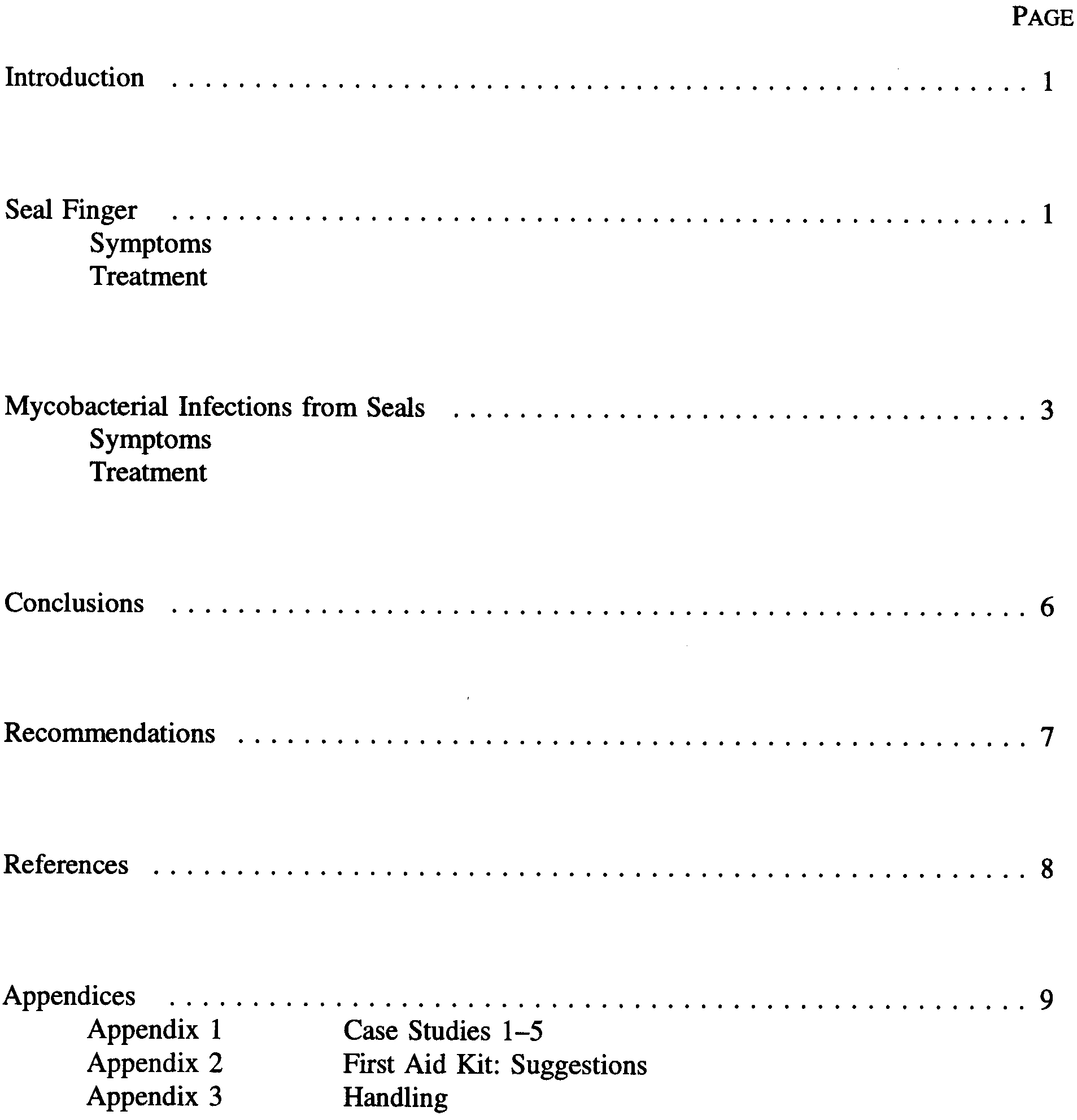 CONTENTS
 APPENDIX 3