Ammonia excretion and urea handling by fish gills: present understanding and future research challenges

JOURNAL OF EXPERIMENTAL ZOOLOGY 293:284–301 (2002) Ammonia Excretion and Urea Handlingby Fish Gills: Present Understandingand Future Research Challenges MICHAEL PATRICK WILKIE*Division of Life Sciences, University of Toronto at Scarborough, Scarborough,Ontario, M1C 1A6 Canada In fresh water fishes, ammonia is excreted across the branchial epithelium via 3 diffusion. This NH3 is subsequently trapped as NH4 in an acidic unstirred boundary layer lying next to the gill, which maintains the blood-to-gill water NH3 partial pressure gradient.
Whole animal, in situ, ultrastructural and molecular approaches suggest that boundary layeracidification results from the hydration of CO2 in the expired gill water, and to a lesser extent H+excretion mediated by apical H+-ATPases. Boundary layer acidification is insignificant in highlybuffered sea water, where ammonia excretion proceeds via NH diffusion due to the greater ionic permeability of marine fish gills. Although Na+/H+ exchangers(NHE) have been isolated in marine fish gills, possible Na+/NH+ evaluation using modern electrophysiological and molecular techniques. Although urea excretion(JUrea) was thought to be via passive diffusion, it is now clear that branchial urea handling requiresspecialized urea transporters. Four urea transporters have been cloned in fishes, including the sharkkidney urea transporter (shUT), which is a facilitated urea transporter similar to the mammalianrenal UT-A2 transporter. Another urea transporter, characterized but not yet cloned, is thebasolateral, Na+ dependent urea antiporter of the dogfish gill, which is essential for urea retention inureosmotic elasmobranchs. In ureotelic teleosts such as the Lake Magadi tilapia and the gulftoadfish, the cloned mtUT and tUT are facilitated urea transporters involved in JUrea. A basolateralurea transporter recently cloned from the gill of the Japanese eel (eUT) may actually be importantfor urea retention during salt water acclimation. A multi-faceted approach, incorporating wholeanimal, histological, biochemical, pharmacological, and molecular techniques is required to learnmore about the location, mechanism of action, and functional significance of urea transporters infishes. J. Exp. Zool. 293:284–301, 2002.
Although the deamination of excess amino acids Since the pK0 of this relationship is approximately liberates carbon skeletons that can be channeled 9.5 (T = 151C; Cameron and Heisler, ’83), more into gluconeogenic pathways or the citric acid than 95 percent of the total ammonia concentra- cycle, this process also leads to the production of highly toxic ammonia (Mommsen and Walsh, ’92; in fishes at physiological pH (e.g., arterial pH of Wood, ’93). In fishes, most ammonia production takes place in the liver, although the enzymes associated with amino acid deamination may be increase as a result of the degradation of organic found in other tissues including the muscle, matter in the sediments of marine and fresh water intestine, and kidney (Mommsen and Walsh, environments, where ammonia buildup may be ’92). Ammonia may also originate in the muscle especially pronounced when nitrification is im- due to the deamination of adenylates in exercising peded as a result of low environmental oxygen fish (Driedzic and Hochachka, ’76), and possibly in concentrations. In addition, ammonia concentra- fish subjected to low environmental O2 concentra- tions may become elevated as a result of crowding In solution, ammonia exists as either un-ionized *Correspondence to: Michael P. Wilkie, Division of Life Sciences, University of Toronto at Scarborough, Scarborough, Ontario, Canada Received 9 April 2002; Accepted 10 April 2002 Published online in Wiley InterScience (www.interscience.wiley.
in fish holding pens or ponds, and from anthro- arises from the catabolism of excess purines pogenic inputs arising from agricultural run-off, through the process of uricolysis. The ornithine sewage, or industrial sources (see Alabaster and urea cycle (OUC), which accounts for the bulk of Lloyd, ’80, for review). Such elevations of environ- urea production in mammals and amphibians mental ammonia may result in histological da- (Wright, ’95), is also active in elasmobranchs mage to the gills of fishes (Smart, ’76) and (Anderson, 2001) and the coelacanth (Latimeria therefore compromise processes such as gas chalumnae; Brown and Brown, ’67), lungfishes exchange, ion regulation, and acid–base regula- (Janssens and Cohen, ’66), and a few selected tion. Ammonia also readily diffuses across the gill teleosts, including the Magadi tilapia (Randall as NH3 under such conditions, but once in the et al., ’89), the gulf toadfish (Walsh, ’97), and the air-breathing Indian catfish (Heteropneustes fossi- lis; Saha and Ratha, ’89). Urea is also produced via rotoxicity (see Cooper and Plum, ’87, for review) the arginase-mediated hydrolysis of dietary argi- characterized by hyperactivity, convulsions, coma, nine. Although trimethylamine oxide and amino and eventually death (Alabaster and Lloyd, ’80).
acids, such as glutamine, may be produced in appreciable quantities by fishes, no studies have metabolism (Arillo et al., ’81) and oxygen delivery conclusively demonstrated that these products to the tissues (Smart, ’78; Arillo et al., ’81). In directly contribute to N-waste excretion (see general, fishes are much more resistant to build- up of internal ammonia than are terrestrial Since ammonia and urea metabolism have been vertebrates, but if blood TAmm exceeds 1.0 extensively reviewed in recent years, readers are mmol Á LÀ1, death results in many fishes (Lumsden asked to consult topical reviews for further details et al., ’93; Knoph and Thorud, ’96).
(e.g., Wood, ’93; Wright, ’95; Walsh, ’97; Anderson, As ammonia is highly toxic, it must either be 2001). The remainder of this article will focus on excreted or be converted to less toxic end- how ammonia and urea are handled by different products, such as urea or uric acid. Uric acid, fishes, with a particular emphasis on the gills, the which is mainly excreted by birds, reptiles, and main site of N-waste excretion in most groups many terrestrial invertebrates, requires little studied to date (Wood, ’93). Efforts will be made to water and does not appear to be excreted in contrast the different strategies fishes use to significant quantities by fishes (Wood, ’93; Wright, excrete their N-wastes in marine and fresh water ’95). Although urea is much less toxic than systems, and to touch on strategies of N-waste ammonia, it is more expensive to produce, requir- excretion that have been observed under more ing at least 2 additional molecules of ATP extreme conditions, such as air exposure or (Mommsen and Walsh, ’91). Although the dipolar prolonged exposure to saline–alkaline environ- nature of urea makes it almost as soluble as ments. As mechanisms of nitrogenous waste ammonia in water (Wood ’93), its low lipid excretion have been reviewed in the last 5–10 solubility (the olive oil–water partition co-efficient years (e.g., Wood, ’93, 2001; Wilkie, ’97; Walsh and is 1.5 Â 10À4 ; Walsh, ’97) suggests that membrane Smith, 2001), I will focus on more recent advances, permeability to urea is at least 2 orders of with particular emphasis on the role that mole- magnitude lower than that of ammonia. It there- cular biology, immunodetection techniques, and fore makes sense that the vast majority of marine ultrastructural analyses have played, and con- and fresh water fishes, including the teleosts and tinue to play, in improving our understanding of lampreys, excrete 80–90% of their nitrogenous how ammonia and urea are handled by the gills of wastes (N-waste) as ammonia and the remainder as urea (Wood, ’93; Wright, ’95). Exceptionsinclude the ureotelic elasmobranchs (Wood et al., ’95a) and unique teleosts such as the gulf toadfish (Opsanus beta; Wood et al., ’95b) and the Lake Magadi tilapia (Alcolapia grahami; formerly Or- The bulk of evidence generated over the last eochromis alcalicus grahami; Randall et al., ’89), 10–20 years indicates that branchial ammonia excretion JAmm in fresh water mainly takes place In most fishes, including larval lampreys (Wilkie down favourable blood-to-water NH3 diffusion et al., ’99) and teleosts (Florkin and Dechateaux gradients (Fig. 1). This strategy is best appreciated ’43; Wright ’93; Wilkie et al., ’93), urea mainly by first considering the physicochemical properties only 0.04–0.08 (Evans and Cameron, ’86). Thus,NH3 lipid solubility is only moderate and muchlower than that of CO2 (Knepper et al., ’89). As thelipid solubility of NH3 is not especially high, doesNH3 enter the lipid bilayer at all during its transitacross the gill epithelium? As pointed out by Wood (’93), one possibility is that NH3 moves through aqueous pores, ratherthan the lipid bilayers. Since the solubility inwater of NH3 is approximately 1,000 times greaterthan that of CO2 and is more than 20,000 timesgreater than that of O2 (Cameron and Heisler, ’83;Boutilier et al., ’84), it should readily move downfavourable PNH through aquaporin 1 (AQP1) expressed in oocytes 
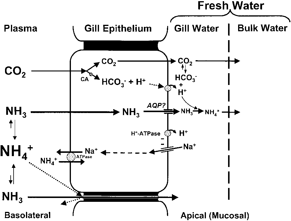
Model of ammonia excretion for fresh water fishes.
of the African clawed frog (Xenopus laevis; Under steady state conditions, CO2 excreted across the gills is Nakhoul et al., 2001). Molecular and electrophy- hydrated in the gill water (unstirred boundary layers) to H+ siological studies examining the possible expres- and HCO3 . Although carbonic anhydrase (CA) is essential forthe hydration of CO sion of aquaporins in fish gill epithelia are still 2 in the cytosol, it is now questionable if gill surface CA plays any role in the hydration of CO2 in the unstirred boundary layers. Nonetheless, H+ generated via measured water flux across elasmobranchs and CO2 hydration and H+-ATPase mediated H+ extrusion acidifies the gills unstirred boundary layers. As a result, The dominance of passive NH3 diffusion in fresh NH3 is trapped as NH4 as it passively diffuses across theapical (mucosal) membrane or passively leaks across the gill water is based on observations that JAmm requires via paracellular routes. This H+ trapping of NH3 ensures that a suitable blood-to-water PNH gradient (Fromm favourable NH3 partial pressure (PNH ) gradients are main- and Gillette, ’68; Maetz, ’72, ’73; Cameron and tained between the gill cytosol and the unstirred boundary Heisler, ’83; Wright and Wood, ’85; Avella and layers under different environmental conditions (e.g., elevated Bornancin, ’89; Wilson et al., ’94). This theory is ammonia or pH). Ammonia likely enters the gill via passiveNH supported by the inhibition of JAmm that results 3 diffusion, and recent studies suggest that a unique Na+ ATPase may also contribute to basolateral when trans-branchial PNH gradients are reversed ammonia transport. It is unlikely that significant NH+ or reduced at high ambient TAmm (Fromm and diffusion takes place in fresh water due to the deep tight Gillette, ’68; Cameron and Heisler, ’83; Wilson junctions that are present between adjacent branchial epithe- et al., ’94) and/or greater water pH (Wright and lial cells. The possible role of aqueous channels (aquaporins; Wood, ’85; Wilkie and Wood, ’91; Yesaki andIwama, ’92; McGeer and Eddy, ’98). Further,moderately lower water pH stimulates JAmm by of ammonia in more detail. Although the majority increasing the blood–water NH3 diffusion gradient in fishes (Maetz, ’72, ’73; Wright and Wood, ’85; above), due to its positive charge it cannot Claiborne and Heisler ’86; Avella and Bornancin, penetrate the lipid phase of cell membranes ’89). In many instances, however, maintenance of (Knepper et al., ’89). In addition, the gills of fresh these trans-branchial PNH gradients relies upon water fish are relatively ‘‘tight’’ to cations (Evans, the hydration of CO2 in the unstirred boundary layers on the apical side of the gill epithelium radius that is slightly larger than Na+ and approximately the same as K+ (Knepper et al., ’89), it is unlikely that appreciable passive NH+ (Fig. 1), which is tied to the hydration of CO2 to diffusion takes place under typical fresh water 3 and H+, is based on early observations by conditions. Further, the electrochemical gradients Lloyd and Herbert (’60), who found that ammonia toxicity is reduced at higher water PCO . Based on are much less than those favouring diffusive Na+ careful measurements of inspired and expired gill and K+ losses. Although NH3 is about 10–1,000 water pH, Wright et al. (’89) later suggested that times more permeable in gill epithelia than NH+ the H+ arising from CO2 hydration traps NH3 as NH4 as it enters the unstirred boundary layers of mucus and water lying next to gill. Although pH water pH. However, if CA were involved in the can drop substantially (0.3–1.5 pH units) as water CO2 hydration reaction, expired gill water pH crosses the gill (Wright et al., ’86; Playle and should equal the theoretical pH that would result Wood, ’89; Lin and Randall, ’90), the extent of 2 were completely hydrated to HCO3 and H+.
acidification is dependent upon on water buffer capacity and inhalant pH (see below).
theoretical pH would constitute a ‘‘disequilibrium Direct evidence of an association between JAmm pH’’ (Gilmour, ’98). Wright et al. (’86) noted a and CO2 excretion in fresh water is demonstrated disequilibrium pH after acetazolamide was added by experiments employing isolated perfused head to the water of their IPHP preparation, suggesting preparations (IPHP). For instance, JAmm is in- that CA catalyzes CO2 hydration in the gill water.
hibited when branchial CO2 excretion is reduced However, Henry and Heming (’98) point out that by perfusing the basolateral side of the prepara- as a strong buffer, acetazolamide addition to the tion with CO2-free saline (Payan and Matty, ’75), water would inhibit boundary layer acidification or by inhibiting intracellular carbonic anhydrase by increasing the water’s non-bicarbonate buffer- (CA) using acetazolamide (Diamox), which inhi- ing capacity, independent of acetazolamide’s af- bits CO2 formation within the gills (Wright et al., fects on CA itself. Thus, reductions in JAmm ’89). However, when water buffer capacity is following acetazolamide addition to the water increased using TRIS, boundary layer acidification (McGeer and Eddy, ’98) are likely artifacts due is prevented and JAmm is reduced (Wright et al., to greater water buffering capacity. As the ’89). As the buffer would bind any H+ arising from uncatalyzed CO2 hydration reaction would likely CO2 hydration in the gill bath, this further be very fast in poorly buffered waters, CA may not demonstrates that a tight coupling between CO2 even be necessary. Indeed, a marked disequili- excretion and JAmm exists in waters of low to brium pH is observed in the expired gill water of moderate buffer capacity. Indeed, this same both the dogfish, Squalus acanthias and rainbow approach can be used to block boundary layer trout, indicating that gill surface CA plays no role acidification in whole fish by adding preparations in CO2 hydration in well-buffered sea water (Perry such as HEPES to the water. For instance, JAmm is initially reversed when rainbow trout (Oncor- Based on these more recent interpretations, it is hynchus mykiss) are exposed to 5 mmol Á LÀ1 questionable if gill surface CA plays any role in boundary layer acidification in fresh water.
gradually recovers as the blood-to-water PNH Although similar approaches to those described gradient is re-established (Wilson et al., ’94). As in sea water (Perry et al., ’99) are needed to boundary layer pH would be identical to the confirm this hypothesis, it is also clear that measured bulk water pH in such experiments, boundary layer acidification may only be impor- manipulations of water NH3 underscore the tant in waters with relatively low buffer capacities.
dependence of JAmm upon the blood-to-gill bound- Indeed, at higher buffer capacity, boundary layer ary layer PNH gradient in fresh water trout acidification and NH3 trapping in the gill water (Wilson et al., ’94). Similarly, measurements of should decrease (Wright et al., ’89; Wilson et al, ammonia and net acid excretion in water buffered ’94; Salama et al., ’99). This is illustrated by the with HEPES reveal that at water pH values Lahontan cutthroat trout, which lives in the ranging from pH 7.7 to 8.2, JAmm declines as the highly buffered waters (titration alkalinity: 23 pH (alkalinity) of the boundary layer water mmol Á LÀ1) of alkaline Pyramid Lake, Nevada (pH increases due to gradual reductions in the blood- 9.4; Wright et al., ’93). Although boundary layer to-water PNH gradient (Salama et al., ’99).
acidification is impossible for this fish, it main- Although boundary layer acidification explains tains favorable blood-to-water PNH gradients by how JAmm persists in the face of apparent inward virtue of its high resting blood pH (pH 8.0) and PNH gradients calculated from bulk water pH and plasma TAmm (Wright et al., ’93; Wilkie et al., ’94).
NH3 measurements (e.g., Wright and Wood, ’85; As ammonia toxicity could be more pronounced Wilkie and Wood, ’91; Yesaki and Iwama, ’92), the when ammonia increases in well-buffered waters, proposed mechanism is controversial. The identi- buffer capacity might be considered when water fication of CA on the external apical surface of the quality criteria for ammonia are drafted or revised gill (Wright et al., ’86; Rahim et al., ’88) suggested this enzyme catalyzes CO2 hydration in the bound- Although appreciable apical Na+/H+ exchange ary gill water, resulting in decreased expired can likely be ruled out in fresh water (see below), evidence that a V-type H+-ATPase is present in accessory cells (Fig. 2; Sardet, ’80). Although the apical epithelium of gill pavement cells (Lin this arrangement substantially increases bran- et al., ’94; Sullivan et al., ’95, ’96) suggests this chial cation (Na+) permeability (Marshall, ’95; transporter also contributes to gill water acidifica- Karnaky, ’98), it is also likely that it provides a tion (Lin and Randall, ’90). Indeed, as this H+- ATPase is closely coupled to channel-mediated Na+ uptake across the gills, it may explain why Recently, a cultured branchial epithelial cell the addition of the Na+ channel blocker amiloride preparation comprised of both chloride cells and to water inhibits JAmm (e.g., Kirschner et al., ’73; pavement cells, and containing high-resistance Payan, ’78; Wright and Wood, ’85; Yesaki and ‘‘tight junctions,’’ exhibited significant NH+ Iwama, ’92; Wilson et al, ’94; McGeer and Eddy, NH3 permeance under fresh water conditions ’98). In such situations, amiloride would not only interfere with Na+ channel access, it would alter significantly correlated with the basolateral-to- apical membrane potential and therefore inhibit electrogenic H+-ATPase activity (Harvey, ’92; across the preparation. Significant basolateral-to- apical NH4 diffusion was also supported by the or moderately buffered waters following amiloride tight relationship between JAmm and the membra- treatment likely reflects decreased boundary layer ne’s electrical conductance, after correcting for acidification resulting from decreased H+-ATPase NH3 diffusion. Although convincing, it is still mediated H+ extrusion. Indeed, when boundary unclear how closely this preparation mimics the layer acidification is impossible in highly buffered true ‘‘in vivo’’ situation as the ammonia concen- waters, amiloride has no affect on JAmm by trations on the basolateral side of the preparation rainbow trout, even in the face of large reductions were relatively high (650 mmol Á LÀ1). Further, (B90%) in Na+ uptake (Wilson et al., ’94).
anatomical factors, such as lamellar blood flow Similarly, JAmm is unaltered in the Lahontancutthroat trout when amiloride is added to thehighly buffered waters of Pyramid Lake (Wrightet al., ’93).
Due to the higher buffer capacity of sea water, and the ‘‘leakiness’’ of the marine fish gill tocations such as NH+ CO2 excretion and JAmm in sea water is unlikely.
As the continual flux of NH+ boundary layers would always result in low NH3, alinkage between JAmm and CO2 would be unne-cessary (Wright et al., ’89). Nonetheless, there islikely significant NH3 diffusion in sea water fishesas demonstrated by the development of a meta-bolic acidosis following NH4Cl infusions in sculpin(Myoxocephalus octodecimspinosus), which likelyresults from rapid losses of NH3 across the gillepithelium (Claiborne and Evans, ’88).
Model of ammonia excretion for marine fishes.
Ammonia excretion in sea water is likely a combination ofpassive NH + diffusion, and to a lesser extent, apical exchange. As in fresh water, NH3 diffusion is dependent upon the presence of suitable PNH gradients between the blood and the water. Passive NH+ diffusion the marine fish gill, but it is unlikely in fresh takes place down favourable electrochemical gradients via shallow (‘‘leaky’’) paracellular tight junctions, while Na+/H+ mmol Á LÀ1; Heisler, ’90) leakage of NH+ exchange proteins (e.g., NHE-2) may provide a route for apical paracellular routes in fresh water fishes is mini- 4 ) exchange. As in fresh water, the possible role of mized by the deep tight junctions between a unique, basolateral Na+ dependent NH4 ATPase deservesinvestigation. However, there is also convincing evidence that adjacent cells in the gill epithelium (Fig. 1; Sardet, enters the gill cytosol by displacing K+ on the ’80). In contrast, marine fishes have shallow tight branchial Na+:2ClÀ:K+ co-transporter and the Na+/K+ junctions between chloride cells and adjacent ATPase. See text for further details.
and water flow across the gills, which could The absence of appreciable acid–base disturbances profoundly influence ammonia delivery to and during high external ammonia exposure also removal from the gill’s microenvironment were demonstrates that the teleost gills have significant not considered. Nor were hormonal factors con- sidered, which could potentially influence bran- Evans, ’88; Wilson and Taylor, ’92). If NH3 entry were dominant under such conditions, a metabolic prolactin is known to reduce branchial ion and alkalosis would arise due to the weakly basic water permeability (Evans, ’84a), little is known properties of NH3. Indeed, TAmm accumulation about how prolactin might affect gill NH+ is greater in sea water- versus fresh water- ability. Nonetheless, these data suggest that the acclimated rainbow trout during ammonia expo- sure (Wilson and Taylor, ’92), which could is not yet resolved and that there is clearly a need make marine fishes more vulnerable to ammonia for a model epithelium that considers hormonal and other factors. As it will be more challenging toincorporate anatomical features into such a model, consideration should be given to additional in vitro models, such as the opercular epithelium of the killifish (Fundulus heteroclitus; Marshall, ’85, change in fresh water fish gills was proposed by ’95), isolated lamellae (Weihrauch et al., ’99), or August Krogh over 60 years ago (Krogh, ’39), and other branchial epithelial preparations.
numerous studies supporting apical Na+/NH+ Both the killifish and isolated lamella prepara- exchange have been published since (see Wilkie, tions would make it possible to isolate ammonia ’97, for review). In this model, Na+ uptake across movements taking place across the basolateral or the apical (mucosal) side of the gill is tied to NH+ apical membrane of the gills, using electrophysio- extrusion, which replaces H+ on an electroneutral logical tools such as the patch clamp or the Ussing Na+/H+ antiport. However, as electroneutral Na+/ chamber. The Ussing chamber would make it possible to determine how changing ammonia or inwardly directed Na+ gradients (Grinstein and hormone concentrations in the blood influences Wieczorek, ’94), the concentration of Na+ in fresh ammonia movements across the basolateral mem- water is insufficient to drive such an antiporter brane in relative isolation from the apical mem- (Potts, ’94; Wilkie, ’97). Recognizing this limita- tion, the most likely arrangement for fresh water conjunction with isolated or cultured branchial Na+ uptake is one in which Na+ moves through epithelial cells, could also be used to determine if apical channels, down favorable electrochemical gradients generated via proton pump-mediated gills. These techniques could also be used to H+ extrusion (Avella and Bornancin, ’89). The determine how transcellular or paracellular am- localization of an electrogenic proton pump (V- monia movements are influenced by alterations in type H+-ATPase) in the apical epithelium of gill pavement cells using immunocytochemistry, Wes- the basolateral or apical membranes of the gill in tern blotting, and in situ hybridization (Lin et al., both marine and fresh water environments.
’94; Sullivan et al., ’95, ’96) supports this more Indeed, the euryhaline nature of the killifish recent model of fresh water Na+ uptake (Perry would make it an ideal model for studying and Fryer, ’97; Marshall, 2002, this issue). It mechanisms of ammonia excretion in both fresh should be noted, however, that Na+/H+ exchange is found on the basolateral membrane, where Na+ electrochemical gradients are sufficient to drive clearly important in sea water where JAmm follows Na+/H+ exchange for intracellular pH regulation toadfish, sculpin, and rainbow trout (Evans, ’82; In view of our present knowledge, the inhibition Goldstein et al., ’82). Further, branchial Na+ and of Na+ influx reported to take place at high 4 permeabilities are similar in toadfish (Evans, external ammonia likely arises from a metabolic alkalosis arising from inward NH3 diffusion and ability during acclimation to low-strength sea water (5%) is accompanied by simultaneous mediated H+ extrusion (Avella and Bornancin, increases in NH3 permeability (Evans et al., ’89).
’89). In contrast, reported increases in Na+ influx demonstration that Na+ influx is tightly coupled NH4Cl in intact fish (Maetz and Garcia-Romeu, to H+ excretion in the hagfish (Myxine glutinosa; ’64; McDonald and Prior, ’88; Wilson et al., ’94) Evans, ’84b), the dogfish shark and the gulf likely result from greater metabolic H+ excretion toadfish (Evans, ’82) supports this hypothesis.
In mammals, Na+/H+ exchange is mediated by (see above). Taking into account the proton pump/ at least six isoforms (NHE-1 to NHE-6) that are Na+ channel model, it is therefore likely that present in numerous tissues, including kidney, greater Na+ influx under these conditions is heart, salivary gland cells, intestine, and brain, linked to the favorable electrochemical gradients and that are essential for processes such as cell that arise from increased H+ extrusion. Indeed volume and acid–base regulation (Ritter et al., IPHPs demonstrate that when perfusate TAmm is 2001). In gills, the presence of a Na+/H+ anti- increased at constant pH, JAmm gradually in- porter was first confirmed in the euryhaline crab creases but Na+ influx does not change (Avella Carcinus maenas, in which a crab NHE cDNA was and Bornancin, ’89). Presumably, Na+ influx cloned by Towle and colleagues (’97). Using a remains constant under these conditions because combined molecular physiology approach, Clai- rates of H+ excretion would be relatively stable borne et al. (’99) recently identified 3 separate NHE isoforms (basolaterally located NHE-1 and b- Although NH3 diffusion clearly dominates in NHE; apically located NHE-2) in the marine long- fresh water, the persistence of the apical Na+/ horned sculpin and the euryhaline killifish using exchange hypothesis is due to the simulta- reverse transcriptase polymerase chain reaction neous reductions in JAmm and Na+ influx observed (RT-PCR) and Northern blotting. The membrane in the presence of amiloride (Wilkie, ’97). As specific distribution of these transporters likely mentioned previously, interpretations based on reflects their specialized roles, with basolateral amiloride induced blockage of JAmm should be NHE-1 and perhaps b-NHE, playing a ‘‘house- reconsidered in light of what is now known about keeping’’ role for intracellular pH regulation (e.g., the linkage between the proton ATPase and Na+ ¨rt and Wood, ’96), and the apical NHE-2 likely influx across the gills. Similarly, the ability of involved in net systemic acid excretion (Edwards many fishes to excrete ammonia against inwardly Because NHE isoforms are present in the gills of ammonia (Fromm and Gillette, ’68; Maetz, ’72, a representative agnathan, elasmobranchs, and ’73; Payan, ’78; Cameron and Heisler, ’83; Wright and Wood, ’85; Heisler, ’90; Wilson et al., ’94) is across the gills? There is clear evidence that NH+ likely tied to boundary layer acidification, not can compete with H+ for exchange sites in many increased Na+ influx. Indeed, the expected 1:1 tissues including the mammalian kidney (Good, stoichiometric reduction in JAmm and Na+ influx ’94) and intestine (Cermak et al., 2000), so breaks down under these conditions (Kerstetter et al., ’70; Kirschner et al., ’73). Although initially seem likely in the marine fish gill. To date McDonald and Milligan (’88) reported that Na+ few experiments have incorporated combined influx and JAmm were coupled in a 1:1 ratio in the molecular and physiological approaches to address brook trout (Salvelinus fontinalis), these observa- such questions, but this will likely change over the tions should be interpreted cautiously because next few years as more cDNA libraries are they were unable to completely saturate the Na+ generated for different fish species.
As with fresh water fishes, early theories regarding Na+/NH4 exchange in marine fishes expected to exhibit saturation kinetics, which to were based on correlating changes in water NH+ my knowledge has not been demonstrated.
and Na+ concentration to JAmm. Unlike the situation in fresh water, however, observed lin- unlikely in the fresh water gill, it could be important in sea water, where external Na+ cannot be explained by boundary layer acidifica- concentrations are sufficient to drive such an tion or altered proton-pump/Na+ channel system antiport. Indeed, Evans (’84b) suggests apical activity (see above). For instance, exposure of Na+/H+ exchange was likely present in the gills dogfish pups and the gulf toadfish to Na+-free of marine fishes prior to their invasion of fresh water leads to lower JAmm, suggesting that this water to facilitate metabolic H+ excretion. The process is at least partially dependent upon Na+/ exchange (Evans, ’82). However, amiloride using some of the in vivo and in vitro approaches has little effect on JAmm in marine fishes (Evans et al., ’79; Evans and More, ’88). Althoughamiloride does inhibit JAmm in toadfish, this effectis thought to be mediated by its inhibitory action on the basolateral Na+/K+ ATPase. Indeed, the amiloride effect on JAmm is abolished when theNa+/K+ ATPase is blocked using ouabain (Evans Although basolateral Na+/H+ exchange is well et al., ’89). Thus, it appears that Na+/NH+ exchange makes little contribution to overall JAmm ’96) and marine fishes (Claiborne et al., ’99), under ‘‘normal’’ conditions in sea water (e.g., salinity 35%, TAmm o 100 mmol Á LÀ1). At higher NHE-1 or b-NHE seems unlikely, as the electro- external ammonia concentrations, however, NH+ motive force for Na+ is directed into the gill cytosol. Although NH4 can substitute for Na+ on tial for counteracting reduced or reversed NH+ basolateral NHEs in selected segments of the electrochemical gradients across the gill in sea renal tubule (Good, ’94) and the rat colon (Cermak et al., 2001), TAmm can be very high in the lumen the air-breathing mudskipper (Periopthalmodon of these tissues. Because extracellular Na+ is more schlosseri) to withstand high environmental am- than 2 orders of magnitude greater than NH+ monia and air exposure (Randall et al., ’99). In this concentrations in fish plasma, it is unlikely that unique burrow dweller, the lamellae are not 4 could outcompete Na+ for access to basolat- appreciably involved in gas exchange or JAmm as they are fused to prevent desiccation of the gill Another possible mode of basolateral ammonia during immersion (Wilson et al., ’99). Instead ammonia is excreted into the Na+ rich water K+ ATPase (Mallery, ’83; Towle and Hlleland, trapped against the gill by these fused lamellae via ’87). As branchial Na+/K+ ATPase activity is relatively low in fresh water versus sea water fishes (Karnaky, ’98), appreciable NH4 transport across the basolateral membrane (see below), via this route seems less likely in fresh water. The allows the mudskipper to excrete ammonia against apparent dominance of NH3 diffusion would also make NH4 transport via this route unnecessary.
both water and in air (Randall et al., ’99).
As branchial NHE expression appears to be recently measured in gill homogenates taken from highly plastic, as demonstrated in the killifish rainbow trout, and monitored in the presence of (Claiborne et al, ’99) and the hagfish (Edwards, 2001), it would be informative to establish if tions, no effects on enzyme activity were observed mRNA or protein expression of branchial NHEs (Salama et al., ’99). Recognizing that Na+ would are altered following exogenous ammonia loading.
have to move against its electrochemical gradient, Salama et al. (’99) instead proposed that a unique, place, then increased internal ammonia, due to either ammonia infusion and/or ammonia expo- cilitates a small, but significant amount of NH+ sure, should lead to compensatory increases in loading into gill cell cytosol of the fresh water apical NHE-2 or NHE-3 expression. Indeed, NHE- trout, with the remainder (uncoupled portion) 2 and/or NHE-3 may be involved in JAmm by the taking place via NH3 diffusion. Thus, the updated air breathing mudskipper P. schlosseri (Wilson model of fresh water ammonia excretion proposed et al., 2000). Although a negative result would not here has ammonia entering the gill cytosol as rule out NHE mediated ammonia excretion in fishes, positive findings would strongly support membrane by NH3 diffusion (Fig. 1). Although such an arrangement in the apical membrane of the challenge of future studies will be to identify marine fish gills. Specific NHE antagonists, such as HOE642 (NHE-1) and S3226 (NHE-3; Cermak could be achieved through functional expression et al., 2001), could subsequently be used to studies using Xenopus oocytes, along with in vitro determine which NHE (if any) is involved in approaches such as isolated basolateral membrane vesicles and/or Ussing chamber set-ups.
glutamine could then be exported for hepatic urea reasonable for fresh water fishes, it is less likely in synthesis, or retained as substrate for intra- branchial urea production (Wood et al., ’95a). As gill entirely via paracellular channels or cross the it is becoming increasingly apparent that the basolateral membrane using alternate methods.
handling of urea by the gills and other organs is far more complex than previously believed, this Na+:2ClÀ:K+ co-transporter expressed on chloride testable hypothesis is certainly worth considering.
cells of sea water fishes, as demonstrated using theloop diuretics furosemide and bumetamide in the mammalian renal tubules (Fig. 2; Good, ’94).
As the furosemide and bumetamide-sensitive Previously, many physiologists believed that Na+:2ClÀ:K+ transporter is found localized to urea readily moved across cell membranes by the basolateral membrane of sea water chloride passive diffusion due to its small size (60 Da). As cells (Wood and Marshall, ’94; Karnaky, ’98), NH+ pointed out by several authors (e.g., Hays et al., excretion via this route is feasible but supporting ’77; Sands et al., ’97; Walsh, ’97), however, urea’s evidence is scant. Using IPHPs, JAmm across dipolar structure and low olive oil–water partition dogfish pup gills is reportedly bumetamide sensi- coefficient (approximately 10À4; Walsh, ’97) pre- tive (Evans and More, ’88), but similar evidence is cludes appreciable urea movement through phos- not found in teleosts such as the toadfish (Evans pholipid bilayers without the aid of highly specialized protein channels or transporters. In- Unlike fresh water fishes, marine fishes have deed, the permeability coefficient of urea in greater overall branchial Na+/K+ ATPase activity, artificial bilayers is only about 4 À 10À6 cm Á secÀ1 which is essential for creating the electrochemical (Walsh, ’97). On the other hand, urea’s dipolar gradients required to facilitate paracellular Na+ structure and high water solubility suggest that it excretion (Wood and Marshall, ’94; Karnaky, ’98).
could readily move through aqueous channels Mallery (’83) first demonstrated that NH+ (aquaporins) in close association with water via replace K+ on branchial Na+/K+ ATPase using ‘‘solvent drag’’ (Sands et al., ’97). As solvent drag toadfish gill homogenates. Later experiments, would require the relative permeabilities of urea revealing that basolateral application of oubain and water to be almost identical, the reflection and K+ inhibits ammonia excretion in toadfish coefficient for urea (surea = 1 À Purea/Pwater) IPHPs (Evans et al., ’89), lends further support to would be expected to be very low (Sands et al., ’97). However, surea for eel and rainbow trout gills are 0.85 and 0.83, respectively, suggesting a Na+/K+ ATPase is seen in the air-breathing relatively low solvent drag potential for urea (Pa mudskipper, which expresses oubain-sensitive transport across the basolateral membrane Homer Smith’s classic studies were not only the of its gills (Randall et al., ’99). Along with apical first to identify the gills as a route of nitrogen excretion in fishes (Smith, ’29, ’36), he also allows this mudskipper to excrete ammonia during correctly predicted that urea retention in elasmo- air exposure or high ambient ammonia.
branchs required highly efficient mechanisms of In dogfish, oubain has no affect on JAmm, renal urea reabsorption (Goldstein and Forster, ’71). It is now clear that urea reabsorption in basolateral Na+/ K+ ATPases in elasmobranch elasmobranchs relies, at least in part, on a gills (Evans and More, ’88). Along with low rates Nielsen et al., ’72), which can be non-competitively to the very low ammonia permeability of the shark inhibited by phloretin (Hays et al., ’77). It was not gill, which is reportedly 22-fold lower than that of until the late 1980s that micropuncture and the rainbow trout (Evans and More, ’88; Wood isolated renal tubule studies revealed that urea et al., ’95a). Another factor is the presence of the transport in the inner medullary collecting duct ammonia ‘‘scavenging’’ enzyme glutamine synthe- (IMCD) of the mammalian kidney was via Na+ tase (GSase) within dogfish branchial epithelial dependent facilitated urea transport (Knepper and cells (Wood et al., ’95a), which would minimize Chou, ’95). In mammals, these transporters con- branchial ammonia losses by trapping ammonia as centrate urea in the inner renal medulla to glutamine in the gill cytosol. The resulting maximize water reabsorption, and also help to minimize swelling or shrinkage of red blood cells passing through the vasa recta (Sands, ’99). In thelast decade, extensive research has revealed that Although elasmobranchs excrete 80–90% of urea movements through these structures are not their total nitrogenous wastes as urea (Wood only mediated by facilitated diffusion but also by et al., ’95a), the shark gill should be designed to active transport (Sands, ’99; Bagnasco, 2000). The minimize urea losses. This is no small challenge, first urea transporter identified and cloned using as blood urea concentrations reportedly range modern molecular approaches was the phloretin- from 260 to 800 mmol N Á LÀ1 in elasmobranchs, sensitive UT-A2-facilitated urea transporter in the resulting in massive blood–water urea diffusion IMCD of the rat kidney (You et al., ’93). A variety gradients, which are at least 2 orders of magnitude of urea transporters have since been identified greater than those of teleosts (Wood, ’93). The low using recombinant DNA techniques and func- urea permeability of elasmobranch gills was first tional expression studies using Xenopus oocytes noted by Boylan (’67), who reported that the (Sands, ’99; Walsh and Smith, 2001). The UT-A diffusional permeability of urea in dogfish gills is family of facilitated urea transporters is presently approximately 7.5 Â 10À8 cm Á secÀ1, which is composed of five isoforms (UT-A1, UT-A2, UT-A3, about 50–100 times less urea-permeable than UT-A4, UT-A5), which differ in their dependence on Na+ and/or their sensitivity to various analogs ¨rt et al. (’98) point out, JUrea would approach such as thiourea or acetamide. The UT-B family, 10,000 mmol Á kgÀ1 Á hrÀ1, about 40 times greater composed of two isoforms, is found in red blood than observed rates, if the urea permeability of the cells, the vasa recta of the kidney, and in the brain dogfish gill were similar to that of the rainbow and testes (Sands, ’99; Bagnasco, 2000). Most trout. In such an instance, urea retention for recently, Smith and Wright (’99) isolated a osmoregulation would be untenable as its produc- phloretin-sensitive facilitated urea transporter in tion would be too costly (2 ATP per urea molecule; the kidneys of dogfish (ShUT), which shares 66% amino acid identity with the rat UT-A2 facilitated Unlike the kidneys, urea clearance via the gills urea transporter and 67% identity with the does not appear to be altered by changes in vasopressin regulated urea transporter in frog external salinity. Although renal urea clearance (Rana esculenta). Incorporation of the cloned increases in the euryhaline little skate (Raja complementary RNA (cRNA) into Xenopus laevis erinacea) acutely exposed to dilute sea water, oocytes confirms the ShUT’s role as a facilitated decreases in branchial JUrea are due to a lower urea transporter (Smith and Wright, ’99).
blood–water diffusion gradient for urea rather Since most fishes are ammonotelic, few studies than changes in branchial urea permeability have examined mechanisms of branchial urea (Payan et al., ’73). In general, increased renal excretion JUrea in fishes. Instead, studies have urea clearance explains the much lower urea focused on how urea is produced and its role in ammonia detoxification during environmental euryhaline elasmobranchs in fresh water (e.g., challenges such as elevated ammonia, highly Thorson et al., ’67; Piermarini and Evans, ’98). It alkaline water or air exposure (Wilkie and Wood, would be informative however, to establish if low ’96; Ip et al., 2001). Further, when possible branchial urea permeability is retained in the carrier mediated urea transport across the gills stenohaline fresh water rays (Potamotrygon sp.) of was examined in the tidepool sculpin, previous the Amazon basin of South America, which have assumptions regarding branchial urea excretion mechanisms appeared correct, as JUrea was mmol Á LÀ1 (Thorson et al., ’67). Nonetheless, with unaffected by urea analogues (e.g., acetamide, the possible exception of the stenohaline fresh methylurea, thiourea) and phloretin (Wright water rays (which cannot survive in sea water), the elasmobranch gill has a low intrinsic urea demonstrate that carrier mediated urea handling permeability, which is unaffected by changes in by the gills is essential in the dogfish (Wood environmental salinity. Until recently, however, et al., ’95a; Fines et al., 2001), gulf toadfish there was little information to explain this low (Wood et al., ’98; Walsh et al., 2000), the tilapia of highly alkaline Lake Magadi (Walsh et al., The first indication that a saturable urea ‘‘back- 2001a), and perhaps the Japanese eel (Anguilla transporter’’ might explain the low branchial urea permeability of elasmobranch gills was based on observations that small elevations in blood urea(15%) resulted in no change in JUrea in dogfish(Wood et al., ’95a). Further, acetamide andthiourea infusions led to increased branchial ureaclearance suggesting that these urea analogueswere competing with urea for binding sites on the‘‘back-transporter.’’ As basolateral application ofphloretin resulted in 2-fold increases in JUreaacross the gills of dogfish IPHPs, it lent furthersupport to the possible presence of an inwardlydirected, basolateral urea transporter (Pa ’98). Interestingly, these observations also ruledout a common route of urea and water movement,as phloretin had no effect on branchial water fluxmeasured using 3H2O. Further evidence favouringa basolateral versus apical location for the ureatransporter was provided by the much higher (14- Model of ammonia and urea handling by the fold) rates of urea efflux to the perfusion medium elasmobranch gill. Although ammonia may be excreted in a (basolateral side) versus the water (apical side) manner that is similar to other marine fishes, branchial NH3losses may be minimized by the presence of glutamine that resulted when urea was removed from each synthetase in the gill cytosol, which promotes glutamine formation from NH3 and glutamate. The resulting glutamine could be exported to the liver, where it enters the ornithine kidney, Smith and Wright (’99) first identified a urea cycle (OUC), and/or be retained in the gill cytosol for homologue to this protein in the elasmobranch gill intra-branchial urea synthesis. A high cholesterol:phospholi-pid ratio in the basolateral membrane, along with a urea back- using Northern analysis, but it is unlikely that transporter(s), minimizes passive urea leakage across the gill.
this facilitated urea transporter is involved in urea This enables elasmobranchs to retain urea in the face of retention. Instead, using isolated basolateral extremely high blood-to-water urea diffusion gradients. The membrane vesicles (BLMV) and 14C-urea, Fines basolateral urea back-transporter appears to be a Na+ et al. (2001) identified a saturable, phloretin- dependent, secondary active transporter, that is inhibited ina non-competitive manner by phloretin, and by oubain sensitive urea antiporter on the basolateral mem- induced decreases in Na+/K+ ATPase activity. As revealed brane of dogfish gill, which is competitively by Northern analysis, a facilitated urea transporter (not inhibited by urea analogues such as N-methylurea shown) may also be expressed to a lesser degree in the and nitrophenylthiourea (NPTU). The inhibition elasmobranch gill, although its precise location and physiolo- of urea transport in the presence of oubain and its gical significance deserves further study. See text for furtherdetails.
stimulation in the presence of ATP also suggestsurea transport is energy dependent. The stimula-tion of urea uptake by the BLMVs with increasingNa+ concentration gradients also suggests thatthis urea transporter is Na+ dependent. Thus, it ammonia permeability by scavenging ammonia appears that urea retention in the dogfish relies on that enters that gill cytosol (see above Wood et al., secondary active transport, in which a Na+:urea ’95a). As suggested by the presence of the ShUT antiporter is energized by the continual removal of homologue, a facilitated UT-A2 like urea trans- Na+ from the gill via basolateral Na+/K+ ATPases porter may also be expressed to a much lesser (Fig. 3). Most interestingly, the very high choles- degree. Although its location needs to be estab- terol to phospholipid ratio reported in the baso- lished, an outwardly directed, facilitated urea lateral membrane is also thought to substantially transport system could be important when the urea ‘‘back-transporter’’ is saturated with urea, As suggested by Fines et al. (2001) it is likely a such as might occur following feeding.
combination of Na+ dependent urea ‘‘back-trans-port’’ and a high basolateral membrane cholester- ol:phospholipid ratio that explains the low ureapermeability of the elasmobranch gill (Fig. 3). In Teleosts are not generally faced with the addition, the presence of glutamine synthetase in challenges faced by ureosmotic animals such as the shark gill epithelium may minimize branchial the elasmobranchs and the coelacanths. In fact ureotelic fishes with a fully functional ornithine which metyrapone is used to block cortisol synth- urea cycle, such as the gulf toadfish (O. beta), the esis (Wood et al., 2001). Rather, drops in cortisol oyster toadfish (O. tau), and the Lake Magadi are likely permissive while the proximate cause tilapia (A. grahami; see Walsh, ’97, and Walsh and remains to be elucidated. Although UT-A2 pro- Smith, 2001, for recent reviews) are faced with the teins in mammals are regulated by vasopressin opposite challenge, a need to get rid of urea. The (Sands, ’99), possible stimulation by arginine gulf toadfish excretes most of its urea in distinct vasotocin (AVT), the teleost equivalent, was ruled pulses 1–3 times per day, while excreting primar- out because circulating AVT was unchanged ily ammonia for the remainder (Wood et al., ’95b).
during natural urea pulse events (Wood et al., Although the ecological relevance of this strategy 2001). These findings contrast those of Perry et al.
remains unclear, it is clear that pulsatile urea (’98), who found that pharmacological doses of excretion only occurs under stressful conditions AVT stimulated urea pulse events in cannulated (e.g., crowding, confinement, air exposure; Walsh gulf toadfish. As future studies are clearly et al., ’90, ’94). As in elasmobranchs, virtually all required to identify the urea pulse trigger, it urea is lost across the gills (Wood et al., ’95b; may be informative to determine how candidate Gilmour et al., ’98), and each pulse is accompanied hormones such as AVT influence branchial urea by marked 30- to 40-fold increases in branchial permeability using in vitro approaches such as isolated gill epithelia or cultured epithelial cell basolateral ‘‘back-transport’’ does not account for preparations. On a larger scale, however, experi- the urea pulse, as JUrea is not altered in Na+-free ments must also establish what functional sig- sea water, or by the presence of competitive urea nificance pulsatile urea excretion has for the transport inhibitors (acetamide, thiourea) during natural pulse events (Wood et al., ’98). When urea The mechanism of urea excretion in the gulf is added to the water during natural ‘‘pulse’’ toadfish may be remarkably similar to that events, systemic urea concentrations increase, hypothesized in the Lake Magadi tilapia, from suggesting that a specific facilitated transport which a facilitated urea transporter (mtUT) cDNA system promotes JUrea in the gulf toadfish.
was also recently cloned (Walsh et al., 2001a). As The recent isolation of a toadfish urea transport pointed out earlier, high rates of urea production protein (tUT) cDNA in the gills of the gulf toadfish and excretion are required to promote nitrogen strongly suggests JUrea is via facilitated urea excretion in Lake Magadi’s extremely alkaline transport (Walsh et al., 2000). Incorporation of waters (pH 10; Randall et al., ’89). As in the gulf tUT cRNA into Xenopus oocytes, confirms that the toadfish, TEM suggests that vesicles emanating tUT is a phloretin sensitive urea co-transporter.
from the Golgi apparatus may progressively However, tUT mRNA expression does not change accumulate urea before merging with the apical during actual urea pulse events, suggesting that membranes of pavement cells to eject their this process is regulated beyond the level of contents to the water (Walsh et al., 2001a). Unlike mRNA. This hypothesis is supported by transmis- the gulf toadfish, branchial urea permeability in sion electron microscopy (TEM) analysis, which the Magadi tilapia is constant and about 5 times reveals that increases in branchial urea perme- greater than that observed during natural urea ability during urea pulses are associated increased pulse events by the gulf toadfish (Walsh et al., vesicular traffic in the apical region of branchial 2000). In both cases, respective tUT and mtUT are pavement cells (Laurent et al., 2001). Together, thought to be located in the membranes of these these findings suggest that the vesicles may vesicles to promote urea loading by both the gradually acquire urea via urea transporters toadfish and Magadi tilapia, although these hy- embedded in their membranes, and then merge potheses also await verification using techniques with the apical membrane to release their urea such as immunohistochemistry or in situ hybridi- contents into the environment. Indeed, TEM zation. As the cDNAs have been cloned for these reveals that the vesicles do appear to get larger putative transporters, it should be possible to prior to a urea pulse event. It is not yet clear, construct the respective antibodies or mRNA however, if urea is actually accumulating in the vesicles, or how this process is mediated.
Taken together, the similar modes of JUrea Although declines in cortisol precede urea toadfish and Lake Magadi tilapia raise the in- pulses, this does not appear to be the direct cause triguing possibility that facilitated urea trans- of a pulse event as illustrated in experiments in port may be widespread in the teleosts. Indeed, 
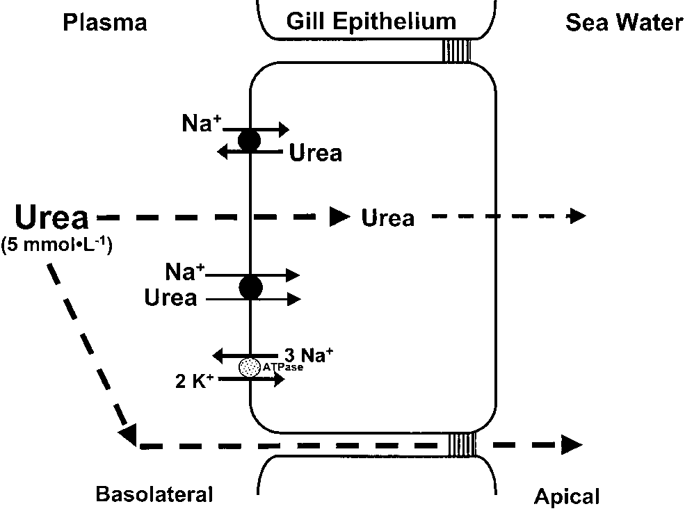
¨rt et al. (’99) suggested that the very low branchial urea permeabilities seen in non-pulsingtoadfish (10À8 cm Á secÀ1) may actually representthe ‘‘true’’ diffusive permeabilities of teleost gills,and that higher branchial urea permeabilities inother fishes (B10À6 cm Á secÀ1) reflect the presenceof moderate numbers of facilitated urea transpor-ters. The recent cloning of a cDNA for an eel(Anguilla japonica) urea transporter (eUT; Mistryet al., 2001) appears to substantiate this hypoth-esis. However, it is not clear if this urea transpor-ter is involved in urea excretion or retention.
Immunohistochemistry, using a polyclonal anti- body raised against the cytoplasmic NH2-terminusof the eUT, indicates that the eUT is located onthe basolateral membrane of branchial chloride Model of urea handling by typical teleosts. In cells. As the physiological properties of the eUT teleosts such as the eels or the tidepool sculpin, urea excretion have not been elucidated, it is not yet clear if it is likely proceeds via passive diffusion across the branchial involved in urea excretion or retention. Mistry epithelium or via ‘‘leaky’’ paracellular channels. Although a et al. (2001) contend that the eUT is a facilitated urea transporter has been isolated on the basolateralmembrane of A. japonica gills, it is unclear if this is a urea transporter involved in JUrea. Northern and Na+:urea antiporter as described in the elasmobranch gill, or Western blot analyses reveal that the eUT is if it is a Na+:urea facilitated transporter. The basolateral markedly up-regulated following sea water accli- location of this protein suggests that it may be a Na+:urea mation. However, branchial urea clearance is antiporter that is involved in urea retention not excretion.
known to decline when related eels, such as the Because the expression of this transporter increases followingsalt water acclimation, when urea excretion decreases in the European eel (Anguilla anguilla), are acclimated related European eel, it seems less likely that this protein is an to sea water (Masoni and Payan, ’74). Although outwardly directed Na+:urea facilitated transporter involved measurements of JUrea are required to confirm in urea excretion. In both cases, basolateral urea transport that A. japonica responds to sea water in a similar would depend upon the continued removal of Na+ via the manner to A. anguilla, it seems more likely that basolateral Na+/K+ ATPase. See text for further discussion.
the greater eUT expression in sea water isassociated with urea retention, not urea excretion(Fig. 4). Clearly, a combination of functional detail in both fresh water and salt water environ- expression studies using Xenopus laevis oocytes ments. Indeed, Walsh et al. (2001b) recently and various physiological approaches (e.g., isolated demonstrated that gill urea transporter mRNA is basolateral membrane vesicles) are required to expressed in the gills of a wide variety of teleosts.
determine if the eUT is a facilitated urea trans-porter involved in JUrea or an active urea trans- basolateral location of the eUT favours the latter In the last 20 years our understanding of hypothesis (see Fines et al., 2001), it raises the ammonia excretion and urea handling by the gills intriguing possibility of increased urea retention of fishes has undergone considerable revision. It is by teleosts in sea water. Indeed, trimethylamine now clear that mechanism(s) of ammonia excre- oxide (TMAO), another nitrogenous osmolyte, is tion in fresh water fishes are vastly different from reportedly higher in certain teleosts following sea those in marine fishes. In fresh water, ammonia is water acclimatization (Van Waarde, ’88). Further, excreted across the branchial epithelium via McDonald and Wood (’98) recently observed active passive NH3 diffusion. This NH3 is subsequently renal urea reabsorption in fresh water-acclimated rainbow trout. Although it is not yet clear what layer lying next to the gill, which ensures that functional significance urea reabsorption would have for fresh water or marine teleosts, it is clear (Fig. 1). On the other hand, boundary layer that urea handling by the gills and kidneys of acidification probably plays no role in marine catadromous (e.g., eel) and anadromous teleosts fishes, in which a combination of passive NH+ (e.g., salmonids) needs to be examined in more and NH3 diffusion likely predominates (Fig. 2).
Differences in urea handling by the gills are less of this and similar techniques for characterizing clear-cut. A basolateral Na+:urea antiporter, along branchial ammonia and urea handling. Ultra- with a high cholesterol:phospholipid ratio, allows structural analyses, using light and electron elasmobranchs to retain urea for the purpose of microscopy, will also be important, especially for osmoregulation in sea water (Fig. 3). The single applications such as immunohistochemistry (e.g., study in which a basolateral urea transporter was Sullivan et al., ’95; Mistry et al., 2001) or in situ isolated in the eel gill raises the possibility that a hybridization (e.g., Sullivan et al., ’96), which will similar antiporter might also be expressed in be essential for isolating transport proteins or teleosts as a means of urea retention, rather than their mRNA, respectively. Of course, molecular excretion (Fig. 4). On the other hand, the cloning techniques will be required to generate the of a Na+ dependent facilitated urea transporter appropriate probes (e.g., polyclonal or monoclonal from the gills of the gulf toadfish and the Lake antibodies, cDNA clones) for such ultrastructural Magadi tilapia, suggests that this protein is analyses. However, molecular techniques will also intricately involved in urea excretion.
be essential for confirming the identity of poten- A limitation of many of the studies examining tial transporters or channels (e.g., aquaporins) ammonia excretion, and to a lesser extent urea through amino acid sequencing and functional handling by the gills, is that research is often expression studies using X. laevis oocytes (e.g., restricted to whole animal or in situ preparations Smith and Wright, ’99; Walsh et al., 2000). In such as isolated perfused heads. Although these many cases, these approaches will also make it approaches have been fruitful, they reveal little possible to identify the regions (e.g., lamellar vs.
about events occurring at the cellular and sub- filamental epithelium) and cell types (e.g., CCs cellular levels of the gill. It is therefore imperative [chloride cells] vs. pavement cells [PVCs]) involved that a model epithelium be developed that allows in ammonia or urea transport. Northern blotting, researchers to identify the mechanisms by which quantitative PCR, and Western blot analysis will ammonia and urea enter and leave branchial also be invaluable for examining transporter epithelial cells. Further use of cultured branchial expression in response to environmental chal- lenges (e.g., salinity, pH, ammonia) or endogenous Kelly and Wood, 2001) should prove very useful, factors (e.g., feeding, hormones). Through this but preparations such as the killifish opercular combined molecular physiology approach, it is epithelium (Marshall, ’85) or isolated gill lamellae probable that many of the questions and hypoth- (Weihrauch et al., ’99) might also be considered, as eses posed in this review will be resolved within they could be used in classical experimental setups such as Ussing chambers. Using these approachesresearchers could separate events occurring in the gill cytosol from those taking place at thebasolateral or apical membrane. For instance, an I express my gratitude to Dr. D.H. Evans for Ussing chamber setup would make it possible to inviting me to contribute to this special edition of examine mechanisms of basolateral ammonia or Journal of Experimental Zoology and for his urea transport through the application of known helpful comments on an earlier draft of this paper.
antagonist or agonists of these processes to the I also thank Dr. C.M. Wood and M.D. McDonald serosal (basolateral side) or mucosal (apical side) for useful discussions regarding some of my solutions bathing the gill membrane preparation.
thoughts on the more controversial aspects of Potential hormonal regulation (e.g., prolactin, ammonia and urea handling by the fish gill.
epinephrine) of ammonia or urea transport couldalso be examined in a similar manner. Of course,modern electrophysiology tools such as microelec- trodes and patch clamps, will also be essential for Alabaster JS, Lloyd R. 1980. Ammonia. Water Quality criteria identifying and characterizing how ammonia and for fresh water fish, 2nd edition. London: Butterworth urea movements take place across the gill using such model epithelia. Because the use of isolated Anderson PM. 2001. Urea and glutamine synthesis: environ- basolateral membrane vesicles has proven invalu- mental influences on nitrogen excretion. In: Wright PA,Anderson PM, editors. Fish physiology, Vol 20: nitrogen able for characterizing how urea is retained by excretion. New York: Academic Press. p 239–277.
elasmobranch gills (Fines et al., 2001), considera- Arillo A, Margiocco C, Melodia F, Mensi P, Schenone G. 1981.
tion should also be given to more widespread use Ammonia toxicity mechanism in fish: studies on rainbow trout (Salmo gairdneri Rich.). Ecotoxicol Environ Safety Evans DH, More KJ, Robbins SL. 1989. Modes of ammonia transport across the gill epithelium of the marine teleost fish Avella M, Bornancin M. 1989. A new analysis of ammonia and Opsanus beta. J Exp Biol 144:339–356.
sodium transport through the gills of fresh water rainbow Fines GA, Ballantyne JS, Wright PA. 2001. Active urea trout (Salmo gairdneri). J Exp Biol 142:155–175.
transport and an unusual basolateral membrane composi- Bagnasco SM. 2000. Urea: new questions about an ancient tion in the gills of a marine elasmobranch. Am J Physiol Boutilier RG, Heming TA, Iwama GK. 1984. Physicochemical Florkin M, Duchateaux G. 1943. Les formes du syste parameters for use in fish respiratory physiology. In: Hoar WS, Randall DJ, editors. Fish physiology, Vol. 10A. New purique chez les animaux. Arch Int Physiol LIII:267–307.
Fromm PO, Gillette JR. 1968. Effect of ambient ammonia on Boylan JW. 1967. Gill permeability in Squalus acanthias.
blood ammonia and nitrogen excretion of rainbow trout In: Gilbert PW, Mathewson RF, Rall DP, editors. Sharks, (Salmo gairdneri). Comp Biochem Physiol 26:887–896.
skates and rays. Baltimore: John Hopkins University Press.
Gilmour KM. 1998. The disequilibrium pH: a tool for the localization of carbonic anhydrase. Comp Biochem Physiol Brown GW Jr, Brown SG. 1967. Urea and its formation in the coelacanth liver. Science 155:570–573.
Gilmour KM, Perry SF, Wood CM, Henry RP, Laurent P, Pa Cameron JN, Heisler N. 1983. Studies of ammonia in the P, Walsh PJ. 1998. Nitrogen excretion and the cardiore- trout: physicochemical parameters, acid–base behaviour and spiratory physiology of the gulf toadfish, Opsanus beta.
respiratory clearance. J Exp Biol 105:107–125.
Cermak R, Lawnitzak C, Scharrer E. 2000. Influence of Goldstein L, Forster RP. 1971. Osmoregulation and urea ammonia on sodium absorption in rat proximal colon.
metabolism in the little skate Raja erinacea. Am J Physiol Pflugers ArchFEur J Physiol 440:619–626.
Claiborne JB, Evans DH. 1988. Ammonia and acid–base Goldstein L, Claiborne JB, Evans DH. 1982. Ammonia balance during high ammonia exposure in a marine teleost excretion by the gills of two marine teleost fish: the (Myoxocephalus octodecimspinosus). J Exp Biol 140:89–105.
4 permeance. J Exp Zool 219:395–397.
Claiborne JB, Heisler N. 1986. Acid–base regulation and ion Good DW. 1994. Ammonium transport by the thick ascending transfers in the carp (Cyprinus carpio): pH compensation limb of Henle’s loop. Annu Rev Physiol 56:623–647.
during graded long- and short-term environmental hyper- Grinstein S, Wieczorek H. 1994. Cation antiports of animal capnia, and the effect of bicarbonate infusion. J Exp Biol plasma membranes. J Exp Biol 196:307–318.
Harvey BJ. 1992. Energization of sodium absorption by the Claiborne JB, Blackston CR, Choe KP, Dawson DC, Harris SP, H+-ATPase pump in mitochondrial-rich cells of frog skin.
Mackenzie LA, Morrison-Shetlar AI. 1999. A mechanism for branchial acid excretion in marine fish: identification of Hays RM, Levine SD, Myers JD, Heinemann HO, Kaplan MA, multiple Na+/H+ antiporter (NHE) isoforms in gills of two Franki N, Berliner H. 1977. Urea transport in the dogfish sea water teleosts. J Exp Biol 315–324.
Cooper AJL, Plum F. 1987. Biochemistry and physiology of Heisler N. 1990. Mechanisms of ammonia elimination in brain ammonia. Physiol Rev 67:440–519.
fishes. In: Truchot JP, Lahlou B, editors. Animal nutrition Driedzic WR, Hochachka PW. 1976. Control of energy and transport processes. 2. Transport, respiration and metabolism in fish white muscle. Am J Physiol 230:579–582.
excretion: comparative and environmental aspects. Com- Edwards SL, Claiborne JB, Morrison-Shetlar AI, Toop T.
parative physiology. Basel: Karger. p 137–151.
2001. Expression of Na+/H+ exchanger mRNA in the gills of Henry RP, Heming TA. 1998. Carbonic anhydrase and the Atlantic hagfish (Myxine glutinosa) in response to respiratory gas exchange. In: Perry SF, Tufts BL, editors.
metabolic acidosis. Comp Biochem Physiol 130A:81–91.
Fish physiology, Vol 17: fish respiration. New York: Evans DH. 1977. Further evidence for Na+/NH+ marine teleost fish. J Exp Biol 70:213–220.
Ip YK, Chew SF, Randall DJ. 2001. Ammonia toxicity, Evans DH. 1982. Mechanisms of acid extrusion by marine tolerance, and excretion. In: Wright PA, Anderson PM, fishes: the teleost, Opsanus beta, and the elasmobranch, editors. Fish physiology, Vol 20: nitrogen excretion. New Squalus acanthias. J Exp Biol 97:289–299.
Evans DH. 1984a. The roles of gill permeability and transport Janssens PA, Cohen PP. 1966. Ornithine–urea cycle enzymes mechanisms in euryhalinity. In: Hoar WS, Randall DJ, in the African lungfish, Protopterus aethiopicus. Science editors. Fish physiology, Vol XB. New York: Academic Press.
Karnaky KJ. 1998. Osmotic and ionic regulation. In: Evans Evans DH. 1984b. Gill Na+/H+ and ClÀ/HCOÀ DH, editor. The physiology of fishes, 2nd edition. Boca systems evolved before the vertebrates entered fresh water.
Kelly SP, Wood CM. 2001. The cultured branchial epithelium Evans DH, Cameron JN. 1986. Gill ammonia transport. J Exp of the rainbow trout as a model for diffusive fluxes of ammonia across the fish gill. J Exp Biol 204:4115–4124.
Evans DH, Kormanik GA, Krasny EJ Jr. 1979. Mechanisms of Kerstetter TH, Kirschner LB, Rafuse D. 1970. On the ammonia and acid excretion by the little skate, Raja mechanisms of ion transport by the irrigated gills of rainbow trout (Salmo gairdneri). J Gen Physiol 56:342–359.
Evans DH, More KJ. 1988. Modes of ammonia transport Kirschner LB, Greenwald L, Kerstetter TH. 1973. Effect of across the gill epithelium of the dogfish pup (Squalus amiloride on sodium transport across body surfaces of fresh acanthias). J Exp Biol 138:375–397.
water animals. Am J Physiol 224:832–837.
Knepper MA, Chou C-L. 1995. Urea and ammonium transport approaches to fish ionic regulation. New York: Academic in the mammalian kidney. In: Walsh PJ, Wright PA, editors.
Nitrogen metabolism and excretion. Boca Raton: CRC Press.
Masoni A, Payan P. 1974. Urea, inulin and para-aminohippu- ric acid (PAH) excretion by the gills of the eel, Anguilla Knepper MA, Packer R, Good DW. 1989. Ammonium trans- anguilla L. Comp Biochem Physiol 47A:1241–1244.
port in the kidney. Physiol Rev 69:179–249.
Mistry AC, Honda S, Hirata T, Kato A, Hirose S. 2001. Eel Knoph MB, Thorud K. 1996. Toxicity of ammonia to Atlantic urea transporter is localized to chloride cells and is salinity salmon (Salmo salar L.) in sea water: effects on plasma dependent. Am J Physiol 281:R1594–R1604.
osmolality, ion, ammonia, urea and glucose levels and Mommsen TP, Walsh PJ. 1991. Urea synthesis in fishes: evolutionary and biochemical perspectives. In: Hochacka P, Mommsen TP, editors. Biochemistry and molecular biology Krogh A. 1939. Osmotic regulation in aquatic animals. New of fishes, Vol 1. New York: Elsevier Science Publications.
York: Dover Publications, Inc. (reprinted 1965).
Laurent P, Wood CM, Wang Y, Perry SF, Gilmour KM, Mommsen TP, Walsh PJ. 1992. Biochemical and environ- ¨rt P, Chevalier C, West M, Walsh PJ. 2001. Intracellular mental perspectives on nitrogen metabolism in fishes.
vesicular trafficking in the gill epithelium of urea-excreting fish. Cell Tissue Res 303:197–210.
Nakhoul NL, Herring-Smith K, Abnulnour-Nakhoul SM, Lin HL, Randall DJ. 1990. The effect of varying water pH on the acidification of expired water in rainbow trout. J Exp expressing aquaporin 1. Am J Physiol 281:F255–F263.
¨rt P, Wood CM. 1996. Na/H exchange in cultured epithelial Lin H, Pfeiffer DC, Wayne Vogl A, Pan J, Randall DJ. 1994.
cells from fish gills. J Comp Physiol 166B:37–45.
Immunolocalization of H+-ATPase in the gill epithelia of ¨rt P, Wright PA, Wood CM. 1998. Urea and water rainbow trout. J Exp Biol 195:169–183.
permeability in dogfish (Squalus acanthias) gills. Comp Lloyd R, Herbert DWM. 1960. The influence of carbon dioxide on the toxicity of un-ionized ammonia to rainbow ¨rt P, Wood CM, Gilmour KM, Perry SF, Laurent P, trout (Salmo gairdneri Richardson). Ann Appl Biol 48: Zadunaiskey J, Walsh PJ. 1999. Urea and water perme- ability in the ureotelic gulf toadfish (Opsanus beta). J Exp Lumsden JS, Wright PA, Derksen J, Byrne PJ, Ferguson HW.
1993. Paralysis in farmed Arctic char (Salvelinus alpinus) associated with ammonia toxicity. Vet Rec 133:422–423.
gill of the perfused head of the trout (Salmo gairdneri).
McDonald DG, Milligan CL. 1988. Sodium transport in the brook trout, Salvelinus fontinalis: effects of prolonged low Payan P, Matty AJ. 1975. The characteristics of ammonia pH exposure in the presence and absence of aluminum. Can excretion by a perfused isolated head of trout (Salmo gairdneri): effect of temperature and CO2-free ringer.
McDonald DG, Prior ET. 1988. Branchial mechanisms of ion and acid–base regulation in the fresh water rainbow trout, Payan P, Goldstein L, Forster RP. 1973. Gills and kidneys in Salmo gairdneri. Can J Zool 66:2699–2708.
ureosmotic regulation in euryhaline skates. Am J Physiol McDonald MD, Wood CM. 1998. Reabsorption of urea by the kidney of the fresh water rainbow trout. Fish Physiol Perry SF, Fryer JN. 1997. Proton pumps in the fish gill and kidney. Fish Physiol Biochem 17:363–369.
McGeer JC, Eddy FB. 1998. Ionic regulation and nitrogenous excretion in rainbow trout exposed to buffered and PJ. 1998. The effects of arginine vasotocin and catechola- unbuffered fresh water of pH 10.5. Physiol Zool 71:179–190.
mines on nitrogen excretion and the cardiorespiratory Maetz J. 1972. Branchial sodium exchange and ammonia physiology of the gulf toadfish, Opsanus beta. J Comp excretion in the goldfish, Carassius auratus. Effects of ammonia loading and temperature changes. J Exp Biol Perry SF, Gilmour KM, Bernier NJ, Wood CM. 1999. Does boundary gill carbonic anhydrase contribute to carbon dioxide excretion: a comparison between dogfish (Squalus movement across the gill of Carassius auratus. J Exp Biol acanthias) and rainbow trout (Oncorhynchus mykiss). J Exp Maetz J, Garcia-Romeu F. 1964. The mechanism of sodium Piermarini PM, Evans DH. 1998. Osmoregulation of the and chloride uptake by the gills of a fresh water fish, Atlantic stingray (Dasyatis sabina) from the fresh water Lake Jesup of the St. John’s River, Florida. Physiol Zool exchanges. J Gen Physiol 47:1209–1227.
Mallery CH. 1983. A carrier enzyme basis for ammonium Playle RC, Wood CM. 1989. Water chemistry changes in the gill microenvironment of rainbow trout: experimental ATPase activity in Opsanus beta. Comp Biochem Physiol observations and theory. J Comp Physiol 159B:527–537.
Potts WTW. 1994. Kinetics of Na+ uptake in fresh water Marshall WS. 1985. Paracellular ion transport in trout animals: a comparison of ion-exchange and proton pump opercular epithelium models osmoregulatory effects of acid hypotheses. Am J Physiol 266:R315–R320.
precipitation. Can J Zool 63:1816–1822.
Rahim SM, Delaunoy J-P, Laurent P. 1988. Identification and Marshall WS. 1995. Transport processes in isolated teleost immunocytochemical localization of two different carbonic epithelia: opercular epithelium and urinary bladder. In: anhydrase isoenzymes in teleostean fish erythrocytes and Wood CM, Shuttleworth TJ, editors. Cellular and molecular gill epithelia. Histochemistry 89:451–459.
Randall DJ, Wood CM, Perry SF, Bergman H, Maloiy GMO, Van Waarde A. 1983. Aerobic and anaerobic ammonia Mommsen TP, Wright PA. 1989. Urea excretion as a production by fish. Comp Biochem Physiol 74B:675–684.
strategy for survival in a fish living in a very alkaline Van Waarde A. 1988. Biochemistry of non-protein nitrogenous compounds in fish including the use of amino acids for Randall DJ, Wilson JM, Peng KW, Kok TWK, Kuah SSL, anaerobic energy production. Comp Biochem Physiol Chew SF, Lam TJ, Ip YK. 1999. The mudskipper, Peri- ophthalmodon schlosseri, actively transports NH+ Walsh PJ. 1997. Evolution and regulation of urea synthesis concentration gradient. Am J Physiol 277:1562–1567.
and ureotely in (Batrachoidid) fishes. Annu Rev Physiol F, Deetjeen P, Paulmichl M. 2001. Na+/H+ exchangers: Walsh PJ, Smith CP. 2001. Urea transport. In: Wright PA, linking osmotic dysequilibrium to modified cell function.
Anderson PM, editors. Fish physiology, Vol 20: nitrogen excretion. New York: Academic Press. p 279–307.
Saha N, Ratha BK. 1989. Comparative study of ureagenesis in Walsh PJ, Danulat E, Mommsen TP. 1990. Variation in urea fresh water, air-breathing teleosts. J Exp Zool 252:1–8.
excretion in the gulf toadfish Opsanus beta. Mar Biol Salama A, Morgan IJ, Wood CM. 1999. The linkage between Na+ uptake and ammonia excretion in rainbow trout: Walsh PJ, Tucker BC, Hopkins TE. 1994. Effects of confine- kinetic analysis, the effects of (NH4)2SO4 and NH4HCO3 ment/crowding on ureogenesis in the gulf toadfish Opsanus infusion and the influence of gill boundary layer pH. J Exp Walsh PJ, Heitz MJ, Campbell CE, Cooper GJ, Medina M, Sands JM. 1999. Regulation of renal urea transporters. J Am Wang YS, Goss GG, Vincek V, Wood CM, Smith CP. 2000.
Molecular characterization of a urea transporter in the Sands JM, Timmer RT, Gunn RB. 1997. Urea transporters in gill of the gulf toadfish (Opsanus beta). J Exp Biol 203: kidney and erythrocytes. Am J Physiol 273:F321–F339.
Sardet C. 1980. Freeze-fracture of the gill epithelium of Walsh PJ, Grosell M, Goss GG, Bergman HL, Bergman AN, euryhaline teleost fish. Am J Physiol 238:R207–R212.
Wilson P, Laurent P, Alper SL, Smith CP, Kamunde C, Schmidt-Nielsen B, Truniger B, Rabinowitz L. 1972. Sodium- Wood CM. 2001a. Physiological and molecular characteriza- linked urea transport by the renal tubule of the spiny tion of urea transport by the gills of the Lake Magadi tilapia dogfish Squalus acanthias. Comp Biochem Physiol 42A: (Alcolapia grahami). J Exp Biol 204:509–520.
Walsh PJ, Wang Y, Campbell CE, De Boeck G, Wood CM.
Smart G. 1976. The effect of ammonia exposure on gill 2001b. Patterns of nitrogenous waste excretion and gill urea structure of the rainbow trout (Salmo gairdneri). J Fish Biol transporter mRNA expression in several species of marine Smart GR. 1978. Investigations of the toxic mechanisms of Weihrauch D, Becker W, Postel U, Luck-Kopp S, Siebers D.
ammonia to fish gas-exchange in rainbow trout (Salmo 1999. Potential active excretion of ammonia in three gairdneri) exposed to acutely lethal concentrations. J Fish different haline species of crabs. J Comp Physiol B 169: Smith HW. 1929. The excretion of ammonia and urea by the Wilkie MP. 1997. Mechanisms of ammonia excretion across gills of fish. J Biol Chem 781:727–742.
fish gills. Comp Biochem Physiol 118A:39–50.
Smith HW. 1936. The retention and physiological role of urea Wilkie MP, Wood CM. 1991. Nitrogenous waste excretion, in the Elasmobranchii. Biol Bull 11:49–82.
acid–base regulation, and ionoregulation in rainbow trout Smith CP, Wright PA. 1999. Molecular characterization of (Oncorhynchus mykiss) exposed to extremely alkaline water.
an elasmobranch urea transporter. Am J Physiol 276: Wilkie MP, Wood CM. 1996. The adaptations of fish to Sullivan GV, Fryer JN, Perry SF. 1995. Immunolocalization of extremely alkaline environments. Comp Biochem Physiol proton pumps (H+-ATPase) in pavement cells of rainbow trout gill. J Exp Biol 198:2619–2629.
Wilkie MP, Wright PA, Iwama GK, Wood CM. 1993. The Sullivan GV, Fryer JN, Perry SF. 1996. Localization of mRNA physiological responses of the Lahontan cutthroat trout for the proton pump (H+-ATPase) and ClÀ/HCOÀ (Oncorhynchus clarki henshawi), a resident of highly alka- in the rainbow trout gill. Can J Zool 74:2095–2103.
line Pyramid Lake (pH 9.4), to challenge at pH 10. J Exp Thorson TB, Cowan CM, Watson DE. 1967. Potamotrygon spp.: Elasmobranch with low urea content. Science 158: Wilkie MP, Wright PA, Iwama GK, Wood CM. 1994. The physiological adaptations of the Lahontan cutthroat trout Towle DW, Hlleland T. 1987. Ammonium ion substitutes for (Oncorhynchus clarki henshawi), following transfer from K+ in ATP-dependent Na+ transport by basolateral mem- fresh water to the highly alkaline waters of Pyramid Lake, brane vesicles. Am J Physiol 252:R479–R489.
Nevada (pH 9.4). Physiol Zool 67:355–380.
Towle DW, Rushton ME, Heidysch D, Magnani JJ, Rose MJ, Wilkie MP, Wang Y, Walsh PJ, Youson JH. 1999. Nitrogenous Amstutz A, Jordan MK, Shearer DW, Wu WS. 1997. Sodium/ waste excretion by the larvae of a phylogenetically ancient proton antiporter in the euryhaline crab Carcinus maenas: vertebrate: the sea lamprey (Petromyzon marinus). Can J molecular cloning, expression and tissue distribution. J Exp Wilson JM, Kok TWK, Randall DJ, Vogl WA, Ip KY. 1999.
United States Environmental Protection Agency. 1999. Up- Fine structure of the gill epithelium of the terrestrial date of ambient water quality criteria for ammonia. EPA- mudskipper, Periophthalmodon schlosseri. Cell Tissue Res 822-R-99-014. Washington DC: USEPA, Office of Water.
Wilson JM, Randall DJ, Donowitz M, Vogl AW, Ip KY. 2000.
Wood CM, Warne JM, Wang Y, McDonald MD, Balment RJ, Immunolocalization of ion-transport proteins to branchial Laurent P, Walsh PJ. 2001. Do circulating plasma AVT and/ epithelium mitochondria-rich cells in the mudskipper or cortisol levels control pulsatile urea excretion in the gulf (Periophthalmodon schlosseri. J Exp Biol 203:2297–2310.
toadfish (Opsanus beta)? Comp Biochem Physiol 129A: Wilson RW, Taylor EW. 1992. Transbranchial ammonia gradients and acid–base responses to high external ammo- Wright PA. 1993. Nitrogen excretion and enzyme pathways nia concentration in rainbow trout (Oncorhynchus mykiss) for ureagenesis in fresh water tilapia (Oreochromis niloti- acclimated to different salinities. J Exp Biol 166:95–112.
Wilson RW, Wright PM, Munger RS, Wood CM. 1994.
Wright PA. 1995. Nitrogen excretion: three end-products, Ammonia excretion in fresh water rainbow trout (Oncor- many physiological roles. J Exp Biol 198:273–281.
hynchus mykiss) and the importance of gill boundary layer Wright PA, Wood CM. 1985. An analysis of branchial acidification: lack of evidence for Na+/NH+ ammonia excretion in the fresh water rainbow trout: effects of environmental pH change and sodium uptake blockade.
Wood CM. 1993. Ammonia and urea metabolism and excre- tion. In: Evans DH, editor. The physiology of fishes. Boca Wright PA, Heming T, Randall DJ. 1986. Downstream changes in water flowing over the gills of rainbow trout.
Wood CM. 2001. Influence of feeding, exercise, and tempera- ture on nitrogen metabolism and excretion. In: Wright PA, Wright PA, Randall DJ, Perry SF. 1989. Fish gill water Anderson PM, editors. Fish physiology, Vol 20: nitrogen boundary layer: a site of linkage between carbon dioxide and excretion. New York: Academic Press. p 201–238.
ammonia excretion. J Comp Physiol 158B:627–635.
Wood CM, Marshall WS. 1994. Ion balance, acid–base Wright PA, Iwama GK, Wood CM. 1993. Ammonia and urea regulation, and chloride cell function in the common excretion in Lahontan cutthroat trout (Oncorhynchus clarki killifish, Fundulus heteroclitus, a euryhaline estuarine henshawi) adapted to highly alkaline Pyramid Lake (pH ¨rt P, Wright PA. 1995a. Ammonia and urea metabolism in relation to gill function and acid–base balance urea excretion in the tidepool sculpin (Oligocottus maculo- in a marine elasmobranch, the spiny dogfish (Squalus sus): sites of excretion, effects of reduced salinity and acanthias). J Exp Biol 198:1545–1558.
mechanisms of urea transport. Fish Physiol Biochem Wood CM, Hopkins TE, Hogstrand C, Walsh PJ. 1995b.
Pulsatile urea excretion in the ureagenic toadfish Opsanus Yesaki TY, Iwama GK. 1992. Some effects of water hardness beta: an analysis of rates and routes. J Exp Biol 198: on survival, acid–base regulation, ion regulation and ammonia excretion in rainbow trout in highly alkaline PJ. 1998. Pulsatile urea excretion in gulf toadfish (Opsanus You G, Smith CP, Kanai Y, Lee W-S, Steizner M, Hediger MA.
beta): evidence for activation of a specific facilitated diffusion 1993. Cloning and characterization of the vasopressin- transport system. J Exp Biol 201:805–817.
regulated urea transporter. Nature 365:844–847.

Source: http://webctupdates.wlu.ca/documents/30184/Wilkie,_MP._2002._Ammonia_excretion_and_urea_handling_by_fish_gills._J._Exp._Zool._293,_284-301.pdf

Microsoft word - treaty of peace israel_jordan_1994.doc

– Politikai, közéleti portál Izrael Államról Treaty of Peace Between the State of Israel and the Hashemite Kingdom of Jordan October 26, 1994 PREAMBLE The Government of the State of Israel and the Government of the Hashemite Kingdom of Jordan: Bearing in mind the Washington Declaration , signed by them on 25th July, 1994, and which they are both committed to honour

Microsoft word - newsletter nejm

This article is reprinted here for the benefit of our clients and their families. We recommend that all of our clients read this article and pass it on to family and friends. Reprinted Article for educational purposes from the New England Journal of Medicine Recently Senator Charles Grassley, ranking Republican on the Senate Finance Committee, has been looking into financial ties between the

Copyright © 2011-2018 Health Abstracts