Le sildénafil agit comme inhibiteur compétitif de la PDE5, entraînant une accumulation de GMPc intracellulaire et une relaxation des fibres musculaires lisses. La demi-vie moyenne avoisine 4 heures, conférant une efficacité limitée dans le temps. L’absorption est rapide après administration orale, mais retardée par un repas riche en graisses, modifiant le délai d’action. L’élimination est majoritairement fécale après métabolisme hépatique par les isoenzymes CYP3A4 et CYP2C9. Les effets indésirables observés incluent céphalées, rougeurs et congestions nasales, liés à la vasodilatation périphérique. Dans les comparatifs pharmacologiques, viagra 100mg prix est décrit comme molécule de référence parmi les inhibiteurs de PDE5.
Vol22_no8_jum_759-.q
Percutaneous Sonographically Guided Treatment of Hydatid Cysts in Sheep Direct Injection of Mebendazole and Albendazole Yahya Paksoy, MD, Kemal Ödev, MD, Mustafa S¸ahin, MD,Bilal Dik, VMD, Recep Ergül, VMD, Ahmet Arslan, PhDObjective. The purpose of this study was to investigate the scolicidal effect of intracystic injection of benzimidazolic solutions in naturally infected sheep with hydatid disease. Methods. Twenty-four sheep with 37 hydatid cysts were included in this study for percutaneous treatment with benzimidazolic solu- tions. The animals were divided into 3 groups: group I, treatment group with mebendazole; group II, treatment group with albendazole; and group III, control group with distilled water. All solutions were given percutaneously under sonographic guidance. Cyst contents were aspirated with a needle, and then scolicidal solutions were injected into the cysts; reaspiration was not done. Routine follow-up sono- graphic images were taken on the 15th day after treatment, then once per month for 3 months, and then at 3-month intervals thereafter. At the 1-month follow-up, the percutaneous aspirate yielded orange juice–like material containing necrotic debris without living scolices. Results. Sonography showed a reduction in cyst size in the benzimidazolic groups (groups I and II) and progressive changes in echo patterns. An anaphylactic reaction was observed during the procedure in 1 animal. After 12 months of sonographic follow-up, the animals in all groups were killed, and macroscopic and micro- scopic changes in tissue samples were evaluated. At autopsy, no cysts with living scolices were found in the benzimidazolic groups, and the appearance of the treated cysts was different from that of those in the control group. Microscopic examination showed the degeneration, necrosis, and thickening of the cyst walls in the treatment groups. Conclusions. Intracystic injection of benzimidazolic solutions as scol- icidal agents may be used for percutaneous treatment of hepatic hydatid cysts in sheep. Key words: benzimidazole; hydatid cyst; percutaneous treatment; scolicidal; sonographic guidance. Abbreviations PAI, puncture, aspiration, and injection Received February 25, 2003, from UltraGörüntüleme Merkezi (private practice), Konya,
ydatid disease is a parasitic infection caused by
Turkey (Y.P.); Departments of Radiology (K.Ö.) and
the larval stage of the tapeworm EchinococcusGeneral Surgery (M.S.), Selcuk University, School ofgranulosus.1 The definitive hosts of E granulosusMedicine, Konya, Turkey; Department of Para-sitology, Veterinary Faculty, Selcuk University, Konya,
are mainly dogs or other carnivores carrying the
Turkey (B.D.); Institute of Veterinary Research,
adult tapeworm in their intestines. Humans and sheep
Konya, Turkey (R.E.); and Department of Medical
(intermediate hosts) are contaminated by ingestion of
Biology and Genetics, Gaziantep University, Schoolof Medicine, Gaziantep, Turkey (A.A.). Revision
parasite eggs, which reach the liver via the portal system to
requested March 12, 2003. Revised manuscript
form hydatid cysts. The disease is endemic in regions such
accepted for publication March 24, 2003.
as the Mediterranean countries and the Middle East. Until
We thank Hüsamettin Vatansev, MD, for helpwith statistical analysis of the data and Evren
recent years, treatment of this disease had been limited to
Burakgazi, MD, for grammatical support.
surgery, but systemic chemotherapeutic agents and percu-
Address correspondence and reprint requests to
taneous treatment have been reported. Drainage of cysts
Yahya Paksoy, MD, Ultra Görüntüleme Merkezi,Serafettin Cad 11, 1-A, Konya 42000, Turkey.
by percutaneous needle puncture is contraindicated
because of potential seeding of living scolices.2–4
2003 by the American Institute of Ultrasound in Medicine • J Ultrasound Med 22:797–803, 2003 • 0278-4297/03/$3.50
Sonographically Guided Treatment of Hydatid Cysts in Sheep
Several types of sclerosing agents, such as
(n = 7 sheep) in group I, 11 cysts (n = 7 sheep) in
group II, and 14 cysts (n = 10 sheep) in group III
hydrogen peroxide, alcohol, and silver nitrate,
are commonly used for percutaneous treatment
The procedure was performed as follows.
of hepatic hydatid cysts.2,5–9 Scolicidal solutions
The cyst was located, and the percutaneous
(e.g., formalin and hypertonic saline) have toxic
approach was selected with the aid of sonogra-
effects on the biliary tree.10–12 Benzimidazole
phy. The maximal cyst diameter was measured,
derivatives (albendazole and mebendazole) are
and the approximate cyst volume was estimated.
the most preferred chemotherapeutic agents,
Prilocaine hydrochloride (Citanest; AstraZeneca
although the efficacy of systemic administra-
International, London, England) was injected at
tion is limited.13–16 When they are taken orally,
the chosen puncture site after sedation with a
only small amounts are transported to the cyst
2% xylazine hydrochloride solution (Rompun,
fluid.17 We thought that direct injection of the
0.1–0.2 ml intramuscularly; Bayer Animal Health,
benzimidazole solutions into the cyst cavities
Monheim, Germany). Puncture was performed
might overcome this obstacle. The aim of this
with 18- to 22-gauge, 13- to 16-cm-long poly-
experiment was to investigate the efficacy of
ethylene needles (Secalon-T; Ohmeda, Swindon,
benzimidazole solutions in the treatment of
England) under sonographic guidance. When the
needle tip was visualized in the correct positionin the cyst cavity, the hydatid fluid was aspirated
Materials and Methods
as much as possible, and the scolicidal solutionwas injected into the cyst. The approximate vol-
ume of the injected solution was equal to half the
research ethics regulations of our institute. We
prior cystic volume. The solution was left in the
screened and selected infected sheep from 475
cyst. Microscopic examination of the aspirates
sheep in herds at the Institute of Veterinary
from the cysts revealed living scolices, laminated
Research in Konya. The sonographic scanning
membranes, hooklets, or combinations thereof
was performed with a real-time gray scale
in all cysts after the first aspirations. The appear-
B-mode scanner and probes of 3.5 MHz (RT-
ance of living daughter vesicles proved the sono-
X200; GE Medical Systems, Milwaukee, WI) and
Routine follow-up sonographic images were
Netherlands). According to the classification of
taken on the 15th day after treatment, then once
per month for 3 months, and then at 3-month
revealed 37 unilocular (type 1) hydatid liver
intervals thereafter. At the 1-month follow-up,
cysts in 24 sheep (8 ewes and 16 White Karaman
cystic contents were reaspirated, and the via-
sheep). Mebendazole tablets (Vermazol, 100 mg;
bility of scolices was examined under light
IE Ulagay, Istanbul, Turkey) were liquefied with
microscopy with eosin Y dye. In each sheep,
isotonic saline, and a solution of 10 g/mL was
only 1 cyst cavity was reaspirated for micro-
prepared. Pure albendazole was obtained as a
scopic examination. Oral antihelmintic agents
solution containing isotonic saline (Andazol,
were not administered to the animals before the
10 g/mL; Biofarma, Istanbul, Turkey). Both solu-
PAI procedure. At the end of the study, the ani-
mals in all 3 groups were given a lethal overdose
Before the procedure, blood samples were taken
of xylazine hydrochloride. Specimens taken from
for serum aspartate aminotransferase, alanine
the groups of animals were examined and com-
aminotransferase, alkaline phosphatase, and
pared microscopically and macroscopically.
total bilirubin levels to test liver functions.
A Kruskal-Wallis test and Mann-Whitney U test
were used for statistical analysis. P < .05 was
group I, treated with mebendazole; group II,
treated with albendazole; and group III, treatedwith distillated water. Group III was kept as a
control group. Thirty-seven cysts from the 24sheep were subjected to echo-guided percuta-
Serial sonographic examinations performed dur-
neous treatment with the puncture, aspiration,
ing the follow-up period of 1 year revealed a
and injection (PAI) technique. There were 12 cysts
gradual decrease in cyst volume and changes in
Paksoy et al
cyst appearance (Figs. 1 and 2 and Table 1). In
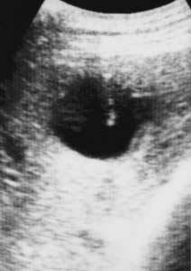
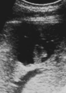
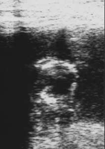
the evaluation of all groups’ posttreatment cystvolumes, the differences between the groupswere significant according to the nonparamet-ric Kruskal-Wallis test (P < .0001). According tothe Mann-Whitney U test, there was a signifi-cant decrease in the cyst volumes in groups Iand II in comparison with group III (P < .0001). There was no significant difference betweenthe cyst volumes in groups I and II (P < .05). Acomparison of pretreatment and posttreat-ment cyst volumes in groups I and II showed asignificant decrease in cyst volumes after treat-ment (P < .002 and .003, respectively). In groupIII, however, there was an increase in cyst vol-umes after PAI, which was statistically signifi-cant (P < .001). Figure 1. Serial sonograms from a sheep with a nonvesicular hepatic hydatid cyst (type I) treated with PAI. A, Liver cyst with well-defined borders and a pure fluid collection during instilla- tion of a benzimidazolic solution. The needle tip is shown in the cystic lesion. B, Follow-up sonogram 1 month after PAI showing collapse of the laminar membrane and disappearance of the anechoic area. C, Follow-up sonogram 6 months after PAI showing obliteration of the cystic cavity by folded membranes (pseudotumor appearance). Sonographically Guided Treatment of Hydatid Cysts in Sheep
Animals were followed up for 12 months after
PAI. In 1 experiment, an anaphylactic reactionoccurred after cyst puncture just before scolicidalagent injection; that animal died and thus wasexcluded from the treatment with scolicidalagents. We considered the death to be due to ananaphylactic reaction caused by cyst contentseeding during cyst puncture, a result of strug-gling by the animal, which did not have adequatesedation. There were no other complicationsduring the procedure.
After PAI, the endocyst separated from the
pericyst in groups I and II. During the follow-up, sonography showed that the cyst cavity wasobliterated by folded membranes and debris(pseudotumor appearance). Living scolices(eosin Y dye) were found in all animals after thefirst aspiration. One month later, cystic con-tents were reaspirated and centrifuged, and theviability of scolices was examined under light
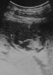
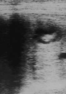
Figure 2. Serial sonograms from a sheep with a type I hydatid cyst. A, Before PAI. B, Sonogram 15 days after PAI showing a partially collapsed cyst cavity. C, One year after PAI, the cyst cav- ity was almost obliterated. Paksoy et al Table 1. Changes in Cyst Volumes in All Groups Cyst Volumes, mL No. of Sheep No. of Cysts
microscopy with eosin Y. One month after percu-
mortality, morbidity, and high recurrence rates.
taneous drainage, no living scolices were
Successful percutaneous treatment of hydatid
observed microscopically in groups I and II; on
liver disease with scolicidal agents also has been
the contrary, living scolices were seen in group
III. These findings imply that both scolicidal
Recently, results of medical treatment with
agents were effective 1 month after percuta-
reported.22–24 Benzimidazole derivatives (fluben-
Because a reduction in cyst size, solidification
dazole, albendazole, and mebendazole) are
of the cyst, absence of a fluid component, and
commonly used for the treatment of hydatid dis-
decreased posterior wall enhancement have
ease in high-risk patients, to prevent secondary
been accepted as healing, as stated in the litera-
hydatidosis, or both.1,2,25,26 However, the results
ture,13,19,20 a postmortem examination of the
of enteric medical therapy are still controversial
entire abdominal cavity was done to check for
and of limited effectiveness.22–24,27–29 Direct cystic
dissemination of the disease. The liver was
injection of these drugs offers the advantage of a
high intracystic drug concentration. Therefore,
microscopically for changes in cyst morphologic
high scolicidal activity can be obtained, and sys-
characteristics. Dissemination of the cysts was
temic side effects can be reduced or avoided. We
not observed in any sheep. The walls of the treat-
hope that pharmaceutical companies can pro-
ed hydatid cysts were hard and solid. Calci-
duce sterile intracystic forms of these agents.
fication of the cyst walls was seen in all but 3
sheep. Microscopic examination of the treated
been treated percutaneously.3,5,6,20,25,26,30,31 The
cysts showed that there were no daughter vesi-
cysts are sterilized with a scolicidal agent, such
cles or living scolices. Hyalinization, thickening,
as hypertonic saline, silver nitrate, 90% alcohol,
and necrosis of the cyst walls were predominant
cetrimide, hydrogen peroxide, benzimidazolic
in the treated cysts. In contrast to those in the
solutions, or formalin.2,3,6,25,32 Unfortunately,
treatment groups, the cysts in the control groups
clinical and experimental studies have shown
had clear fluid with living scolices and thin,
that intracystic injection of scolicidal agents
intact walls. Microscopic examination did not
may cause sclerosing chemical cholangitis.33,34
show any evidence of hepatobiliary toxicity. Liver
Moreover, 1 report described fatal cholangi-
function test results were within the normal
tis.35 Unlike other scolicidal agents, albenda-
zole is not toxic to the liver and biliarystructures at the applied concentration, which
Discussion
potentially decreases the possibility of chemi-cal sclerosing cholangitis.13 Erzurumlu et al10
Hydatid disease caused by E granulosus is a com-
reported that direct application of albendazole
and mebendazole solutions to the biliary sys-
Turkey, and other Mediterranean countries. The
tem of rabbits did not cause any side effects.
conventional treatment of hydatid liver disease
Our study did not reveal any injury to the
is surgery, which is associated with considerable
Sonographically Guided Treatment of Hydatid Cysts in Sheep
In the course of follow-up, sonographically
Ödev K, Paksoy Y, Arslan A, et al. Sonographically
guided reaspiration was performed on 6 cysts in
guided percutaneous treatment of hepatic hydatid
each group. There were no living scolices in the
cysts: long-term results. J Clin Ultrasound 2000;
treated cysts of groups I and II. Living scolices
were found in the control group. Serial sono-
Aygün E, S¸ahin M, Ödev K, et al. The management
graphic examinations over 1 year revealed a
of liver hydatid cysts by percutaneous drainage. Can
marked reduction in lesion size in the treated
groups but not in the control group. A solid pat-tern or a heterogeneous echo pattern was seen in
Ödev K, Aygün E, Kartal A, et al. Percutaneous treat-
the cysts of all animals treated with albendazole
ment of hydatid disease. Turk J Intervent Radiol 1997;
or mebendazole. Postmortem histopathologic
examination of the cysts showed thickening of
10. Erzurumlu K, Özdemir M, Mihmanlı M, Çevikbap U.
the cyst walls, hyalinization, necrosis of the walls,
The effect of intra-operative mebendazole-albenda-
and local calcification. There was no evidence of
zole application on the hepatobiliary system. Eur Surg
biliary toxicity in the liver, such as ductal epithe-
lial proliferation, ductal dilatation, or fibrosis. Inthis study, we observed that there was a correla-
11. Belghiti J, Benhamou TB, Houry S, Grenier P, Hugucer
tion between the follow-up sonographic appear-
M, Fekete F. Caustic sclerosing cholangitis. Arch Surg
We conclude that sonographically guided intra-
12. Polo JR, Garcia-Sabrido JL. Sclerosing cholangitis
cystic injection of benzimidazolic solutions is
associated with hydatid liver disease. Arch Surg 1989;
effective in the treatment of hepatic hydatid cysts
in sheep. In experimental models, intracysticinjection of benzimidazolic solutions as scolici-
13. Deg˘er E, Hökelek M, Deg˘er BA, Tutar E, Asil M,
dal agents should be considered for treating hep-
Pakdemirli E. A new therapeutic approach for the
atic hydatid cysts. Well-designed human studies
treatment of cystic echinococcosis: percutaneous
are needed to establish the efficacy of the PAI
albendazole sulfoxide injection without reaspiration.
Am J Gastroenterol 2000; 95:248–254.
14. Horton RJ. Chemotherapy of Echinococcus infection
References
in man with albendazole. Trans R Soc Trop Med Hyg1989; 83:97–102.
Bezzi M, Teggi A, De Rosa F, et al. Abdominal hydatiddisease: US findings during medical treatment.
15. Ammann RW, Eckert J. Cestodes: Echinococcus.
Gastroenterol Clin North Am 1996; 25:655–689.
Khuroo MS, Zargar SA, Mahajan R. Echinococcus
16. Horton RJ. Albendazole in treatment of human cystic
granulosus cysts in the liver: management with per-
echinococcosis: 12 years of experience. Acta Trop
cutaneous drainage. Radiology 1991; 180:141–145.
Mueller PR, Dawson SL, Ferrucci JT, Nardi GL. Hepatic
17. Erzurumlu K, S¸ahin M, Selçuk MB, Yıldız C, Kerim M.
echinococcal cyst: successful percutaneous drainage.
Intracystic application of mebendazole solution in the
treatment of liver hydatid disease. Eur Surg Res 1996;28:466–470.
Livraghi T, Bosoni A, Giordano F, Lai N, Vettori C. Diagnosis of hydatid cyst by percutaneous aspiration:
18. Gharbi HS, Hassine W, Brauner MW, Dupuch K.
value of electrolyte determinations. J Clin Ultrasound
Ultrasound examination of the hydatic liver.
Filice C, Pirola F, Brunetti E, Dughetti S, Stroselli M,
19. Akhan O, Dinçer A, Gököz A, et al. Percutaneous
Foglieni CS. A new therapeutic approach for hydatid
treatment of abdominal hydatid cyst with hypertonic
liver cysts. Gastroenterology 1990; 98:1366–1368.
saline and alcohol: an experimental study in sheep. Invest Radiol 1993; 28:121–127.
Filice C, Stroselli M, Brunetti E. Percutaneousdrainage of hydatid liver cysts. Radiology 1992; 184:579–580. Paksoy et al
20. Akhan O, Özmen MN, Dinçer A, Sayek F, Göçmen A.
in sheep. Paper presented at: Sixth International
Liver hydatid disease: long-term results of percuta-
Congress of the Mediterranean and African Society
neous treatment. Radiology 1996; 198:256–264.
of Ultrasound International Postgraduate Course ofUltrasound; April 25–28, 1998; Kusadasi, Turkey.
21. Filice C, Di Perri G, Stroselli M, et al. Parasitologic
findings in percutaneous drainage of human hydatid
33. Yılmaz Z, Pekrü I, Sözüer E, Kahya HA, Ye¸silkaya A,
liver cysts. J Infect Dis 1990; 161:1290–1295.
Bengisu N. Sclerosing cholangitis induced by scolici-dal agents injected into the biliary tree of rabbits.
22. Davis A, Pawlowski ZS, Dixon H. Multicentre clinical
trials of benzimidazole carbamates in humanechinococcosis. Bull World Health Organ 1986; 64:
34. Bastid C, Azar C, Doyer M, Sahel T. Percutaneous
treatment of hydatid cysts under sonographic guid-ance. Dig Dis Sci 1994; 39:1576–1580.
23. Todorov T, Vutova K, Mechkov G, Petkov D,
Nedelkov D, Tonchev Z. Evaluation of response to
35. Abbas A, Shadmehr B, Ghaffarinejat MH, Shishineh
chemotherapy of human cystic echinococcosis. Br J
P, Saidi F. Scolicidal agents can cause sclerosing
cholangitis [abstract]. HPB Surg 1990; 2(Suppl):157.
24. Morris DL, Dykes PW, Dickson B, Marriner SE, Bogan
JA, Burrows FG. Albendazole in hydatid disease. BrMed J (Clin Res Ed) 1983; 286:103–104.
25. Gargouri M, Ben Amour N, Chehida FB, et al.
Percutaneous treatment of hydatid cysts(Echinococcus granulosus). Cardiovasc InterventRadiol 1990; 13:169–173.
26. Mente¸s A. Hydatid liver disease: a perspective in
treatment. Dig Dis 1994; 12:150–160.
27. Ammann DW. Improvement of liner resectional ther-
apy by adjuvant chemotherapy in alveolar hydatiddisease. Swiss Echinococcus Study Group (SESG). Parasitol Res 1991; 7:290–291.
28. Davis A, Dixon H, Pawlowski ZS. Multicentre clinical
trials of benzimidazole carbamates in human cysticechinococcus (phase 2). Bull World Health Organ1989; 67:503–508.
29. World Health Organization. Guidelines for treatment
of cystic and alveolar echinococcosis in humans. BullWorld Health Organ 1996; 74:231–242.
30. Khuroro MS, Dar MY, Yattoo GN, et al. Percutaneous
drainage versus albendazole therapy in hepatichydatidosis: a prospective randomized study. Gastro-enterology 1993; 104:1452–1459.
31. Saremi F, McNamara TO. Hydatid cysts of the liver:
long-term result of percutaneous treatment using acutting instrument. AJR Am J Roentgenol 1995; 165:1163–1167.
32. Ödev K, Paksoy Y, Arslan A, Baykan M, Ergül R, Dik
B. Percutaneous treatment of hepatic hydatid cystwith benzimidazolic solutions: an experimental study
Nations conference was first enacted in 1999 and has ratified, in various versions, by all fifty US States, define, Does E-mail Have the Same “that a record or signature may not be denied legal effect Evidentiary Foundation as or enforceability solely because it is in electronic form.” Paper-and-Post Mail? UETA goes further, affirmatively stating that “if a law requires a re
Sisters come to help mother after father's death. LearnThe story of Charlie's freshman year of high school:finding new friends, love, pain, parties and familydrama. A thoughtful look at growing up and makingWriter, American, and Jewish immigrant tell storiesShort stories set in small town Americana in 1943 withStory of survival in Colorado after a flu pandemicBaseball writer covering opening
 Paksoy et al
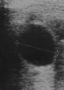
 Sonographically Guided Treatment of Hydatid Cysts in Sheep