Correlation between motor and phosphene thresholds: a transcranial magnetic stimulation study

r Human Brain Mapping 29:662–670 (2008) r
Correlation Between Motor and Phosphene
Thresholds: A Transcranial Magnetic Stimulation Study
Choi Deblieck,1 Benjamin Thompson,2 Marco Iacoboni,1,3,4
and Allan D. Wu1,5*
1Ahmanson-Lovelace Brain Mapping Center, UCLA, Los Angeles, California 2Department of Psychology, UCLA, Los Angeles, California 3Department of Psychiatry and Biobehavioral Sciences, David Geffen School of Medicine at UCLA, 4Brain Research Institute, David Geffen School of Medicine at UCLA, Los Angeles, California 5Department of Neurology, David Geffen School of Medicine at UCLA, Los Angeles, California Abstract: Transcranial magnetic stimulation (TMS) has become a common tool for the brain mapping of awide variety of cognitive functions. Because TMS over cortical regions of interest other than motor cortexoften does not produce easily observable effects, the ability to calibrate TMS intensity for stimulation overnonmotor regions can be problematic. Previous studies reported no correlation between motor thresholds(MT) over the motor cortex and phosphene thresholds (PT) over the visual cortex. However, differentthresholding methods, lighting, and eye-closure conditions were used to determine MT and PT. We investi-gated the correlation between resting MT (rMT), active MT (aMT), and PT in 27 dark-adapted healthy vol-unteers. All thresholds were measured with eyes-open in the dark and determined by gradually reducingstimulation intensity downward. All subjects had aMT and rMT; 21 subjects had measurable PT. rMT was70.4% 6 9.8% (mean 6 SD of maximum stimulator output); aMT was 61.1% 6 7.9%; PT was 82.2% 610.1%. A significant positive correlation was found between aMT and PT (r ¼ 0.53; P ¼ 0.014) with a trendtoward correlation between rMT and PT (r ¼ 0.43; P ¼ 0.052). Our results suggest that sensitivity to TMSover visual and motor cortices may be correlated under similar thresholding procedures. They also providea rationale for the use of easily obtained aMT to calibrate TMS intensities in brain mapping studies thatemploy TMS in cortical regions besides motor cortex. Hum Brain Mapp 29:662–670, 2008.
C 2007 Wiley-Liss, Inc.
Key words: motor threshold (MT); phosphene threshold (PT); cortical excitability; transcranial magnetic Contract grant sponsor: NINDS, NIH; Contract grant number: *Correspondence to: Allan D. Wu, Department of Neurology, K23-NS045764; Contract grant sponsor: NCRR, NIH; Contract David Geffen School of Medicine at UCLA, 710 Westwood Plaza, grant numbers: RR12169, RR13642, RR00865; For generous sup- Reed Bldg A-153, Los Angeles, CA 90095-1769.
port, we thank: Brain Mapping Medical Research Organization, Brain Mapping Support Foundation, Pierson-Lovelace Foundation, Received for publication 29 January 2007; Revised 23 April 2007; The Ahmanson Foundation, William M. and Linda R. Dietel Phi- lanthropic Fund at the Northern Piedmont Community Founda- tion, Tamkin Foundation, Jennifer Jones-Simon Foundation, Capi- Published online 27 June 2007 in Wiley InterScience (www.
tal Group Companies Charitable Foundation, Robson Family and C 2007 Wiley-Liss, Inc.
r Correlation Between Motor and Phosphene Thresholds r
INTRODUCTION
potentially increases the variance of TMS effects, reducesstatistical power of TMS studies, and represents a major li- Transcranial magnetic stimulation (TMS) is a noninva- mitation when considering applications of TMS as a brain sive method for brain stimulation that has become an im- mapping tool across multiple cortical regions [Robertson portant modality for mapping brain–behavior relationships et al., 2003]. The assumption that a relevant proportion of in cognitive neuroscience [Robertson et al., 2003]. In con- TMS threshold measures between different neocortical trast to correlational neuroimaging methods, such as fMRI regions could reflect a shared component of within-indi- or PET, TMS interacts with ongoing brain activity around vidual responsiveness to TMS has been criticized by previ- the region of cortex where induced current under the coil ous studies that demonstrated no correlation between MT is produced. Consequently, TMS can be used to directly and PT [Antal et al., 2003b; Boroojerdi et al., 2002; Gerwig evaluate the critical and causal significance of the stimu- et al., 2003; Stewart et al., 2001b].
lated areas. In conjunction with behavioral tasks, TMS However, none of those studies used similar methods to studies can directly demonstrate causal relationships determine both thresholds (see Table I). For example, while between brain areas and tasks. For example, TMS over the PT is usually measured under dark-adapted conditions, MT occipital lobe can disrupt Braille reading in congenitally is not. The approach toward threshold (upwards, down- blind individuals [Cohen et al., 1997], TMS over Broca’s wards, or variable) is not consistent between or within stud- area can interfere with imitation of hand postures [Heiser ies [Antal et al., 2003b; Boroojerdi et al., 2002; Gerwig et al., et al., 2003], and TMS over the posterior parietal cortex can 2003; Stewart et al., 2001b]. Furthermore, other factors may disrupt feedforward error correction in visually guided have led to additional confounds in previous studies. For example, paired pulses of TMS, which enhance phosphene TMS effects depend on factors, such as cortical target, perception, have been used over the visual cortex in contrast TMS coil geometry, pulse waveform, and stimulation pa- to single TMS pulses over the motor cortex [Antal et al., rameters, such as intensity, frequency, and number of 2003b; Boroojerdi et al. 2002] or different machines were pulses. While the nature of the brain mapping experiment used for PT and MT determinations [Antal et al., 2003b].
can determine cortical targets of interest, the choice of stimu- Since MTs can be affected by these factors [Leon-Sarmiento lation parameters is not always straightforward. Even if all et al., 2005; Tranulis et al., 2006], the possibility exists that technical factors that determine the topography and strength these different methodological factors could have limited of the magnetic field are identical, individual differences in the detection of a correlation between MT and PT.
the intrinsic responsiveness, or excitability, of each subject’s The aim of the present study was to investigate the rela- brain to stimulation will introduce unwanted variability in tionship between MT and PT with psychophysically simi- TMS effects [Robertson et al., 2003]. To assure comparability lar thresholding procedures over motor and visual cortices.
between experimental conditions, TMS intensities should be Specifically, we sought to determine all thresholds under ideally calibrated such that TMS pulses produce a constant both dark-adapted conditions and a uniform systematic neurophysiological effect across subjects.
downward approach toward threshold with single TMS In addition to being variable across subjects, excitability pulses from the same TMS coil. A finding of a significant also varies across different cortical regions and in different MT and PT correlation would suggest some level of com- contexts within a given subject [Robertson et al., 2003].
mon excitability across these two cortical regions. In addi- Over the motor cortex, excitability can be quantified using tion, a significant result would provide a rationale to the a measurable motor evoked potential response (MEP) in a practice of calibrating TMS intensities over different corti- contralateral muscle. The motor threshold (MT) is com- monly defined as the minimum TMS intensity that elicits aMEP above a minimal size and is routinely determined ineach individual subject prior to an experiment. Experimen- SUBJECTS AND METHODS
tal stimulation intensities can then be set at a percentage Subjects
of this MT, which assures that a suprathreshold TMS in-tensity used over the motor cortex in one subject will be We recruited and obtained informed consent from 27 equivalently suprathreshold for another. Over the occipital healthy subjects excluding those with a previous history of cortex, excitability can be assessed using TMS-induced neurological or psychiatric disorders, who did not take phosphenes as a region-specific response measure. Analo- any regular medications, and who did not have exclusions gous to MT, a phosphene threshold (PT) can be defined as relevant to TMS. Study procedures were approved by the the minimum TMS intensity that elicits perception of phos- UCLA Medical Institutional Review Board.
phenes. The PT then becomes a valid reference intensityfor TMS studies of visual perception [Boroojerdi et al., TMS Procedures
2002; Gothe et al., 2002; Hotson and Anand, 1999;Kammer, 1999; Stewart et al., 2001a].
All subjects were dark-adapted for the study by donning The uncertainty in calibrating intensities across subjects lightproof goggles at the start of the experimental session.
for brain regions beyond the motor and visual cortices Goggles were adjusted to ensure that no light was visible.
r Deblieck et al. r
Goggles were designed not to produce pressure on eyelidsand preserve normal blinking. The room was darkened.
Participants were continually reminded to keep eyes open and to fixate forward throughout each thresholding proce- dure. Since different examiners may constitute a significant source of variability [Chaudry et al., 1991], we had the same two investigators present at every session who supervised a consistent application of methods. Each in-vestigator conducted the same thresholding procedure throughout this study. One investigator performed all motor thresholding procedures. A second investigator was only interested in measuring PT for a vision study that required participants to see phosphenes, and performed allsubsequent phosphene thresholding procedures. The sec- ond investigator was blinded to the purpose of this study since no correlation was expected to be found. In addition, the thresholding procedures were spread out over thecourse of several weeks, and no correlation analysis was computed until all subjects for the vision study were Thresholding procedures were conducted on the same day in the following order: (1) measurement of resting motor threshold (rMT), (2) active motor threshold (aMT), and (3) phosphene threshold (PT). Goggles were worn for at least 15 min before rMT thresholding procedures began.
Significant
Adaptation to dark was present for 45 min by the time PT procedures began. To ensure parallel procedures, all thresholds were measured by consistently starting from a clearly suprathreshold intensity and gradually reducing stimulation intensity in steps of 1% until threshold inten- Reporting
sity was obtained. No thresholds were approached from below. Participants wore a tight lycra cap, on which grids were drawn over the region of the left primary motor cor- tex and left occipital visual areas.
We used a Magstim SuperRapid biphasic stimulator with a figure-8 coil (14 cm width) for all motor and PT.
All following thresholds are expressed in percent of maxi- mum stimulator output (MSO) (peak field strength 2 T).
elational
Motor Thresholding Procedures
For rMT and aMT, the figure-8 coil was held tangen- tially to the skull and mediolaterally with the handle pointing backwards and at a 458 angle from the sagittal midline [Brasil-Neto et al., 1992]. Thus, the induced cur- rent pointed forward in a roughly perpendicular manner to the fictitious line of the central sulcus.
Surface EMG electrodes were placed over the right first dorsal interosseus (FDI) muscle. EMG was sampled at 1 kHZ, amplified, and 1-1 kHz bandpass filtered. MEP sizes were measured as peak-to-peak amplitudes. Aliasing of higher frequency components in the EMG signal, where power is minimal, is unlikely to affect thresholding results.
To measure rMT, single pulses of TMS were delivered over the left motor cortex, while the right FDI was kept relaxed. Trials where baseline EMG, in an interval 100 ms prior to TMS pulse, showed visible EMG activity (>20 lV) r Correlation Between Motor and Phosphene Thresholds r
were discarded. TMS pulses were delivered, while the coil close to parameters that would induce reproducible phos- was moved systematically, first at and then, between grid phenes. Positive responses were then qualified as either points. The location that evoked the largest and most reli- central or peripheral in nature. The quality of the visual able MEP amplitudes was designated the motor hotspot.
phenomena was further assessed with open questions (tex- Starting suprathreshold intensities induced clearly dis- tured, colored, shaped and so on). Candidate phosphenes tinguishable MEP’s with every TMS pulse. Intensities were were validated by moving the coil laterally to ensure that then lowered by 1% decrements. The lowest intensity with the perceived phosphene shifted location in a predictable the coil at the motor hotspot for which peak-to-peak MEP manner, validating the retinotopic nature of the visual per- amplitudes greater than 50 lV occurred in at least 5 out of cept. The location that evoked the brightest and most reli- able phosphene was designated the visual hotspot. Then, For aMT, subjects were asked to squeeze a small cylin- from the suprathreshold intensity, intensities were lowered der with a light steady pinch grip, while FDI activation by 1% increments. The lowest stimulation intensity at was monitored online, using EMG to ensure a constant av- which stable phosphenes were perceived in at least 5 of 10 erage level of activity around 100 lV. With the coil held stimulations was recorded as the PT.
over the same hotspot, TMS intensity was then lowered by TMS-induced phosphene perception can be improved by 1% increments from rMT. The lowest intensity for which training in individuals over time or by a period of dark peak-to-peak MEPs greater than 100 lV above baseline adaptation [Boroojerdi et al., 2000a]. Most studies using EMG occurred in 5 out of 10 trials was designated the phosphenes for calibration purposes have not typically aMT. Throughout rMT and aMT procedures, subjects were trained participants to see phosphenes before a threshold- frequently asked to keep their eyes open, while looking ing session, however our phosphene screening procedure gave multiple opportunities for subjects to become familiarwith the concept and appearance of TMS induced phos- Phosphene Thresholding Procedures
phenes. We dark-adapted subjects for 45 min, duringwhich we were able to record motor thresholds on all sub- All PT procedures were done with the FDI muscles jects, while increasing chances of reliable phosphene detec- relaxed. To elicit phosphenes, the coil was positioned tion. Also, because we approached PT by decreasing TMS with the handle pointing upwards, parallel to the sub- intensities, a phosphene was reported on every trial for the ject’s spine [Antal et al., 2003b; Boroojerdi et al., 2002; early part of thresholding procedures allowing participants Stewart et al., 2001b]. The initial position of the coil was to recognize a reliable precept of phosphenes before deter- midline, 2 cm above the inion. Single pulses of TMS were mining PT. This descending approach to phosphene delivered over occipital cortex, while the coil was moved threshold was selected to reduce the risk of participants over a 1 3 1 cm2 interval grid marked on the lycra cap.
having artificially high thresholds as they were not waiting The coil was moved systematically over the left visual for a phosphene to appear, but rather for one that they cortex to induce the perception of phosphenes in the right visual field. All phosphene percepts were initiallydetermined at 100% MSO. The phosphene localization Data Analysis
procedure was designed to maximize the likelihood thatreliable phosphenes were detected: during our first pass rMT, aMT, and PT were compared using one-way over the grid, participants were asked to attend to the repeated measures ANOVA with least significant differ- whole visual field and report the presence or absence of ence post-hoc contrasts. Pearson correlation coefficients any induced visual phenomena after each TMS pulse. If were computed for each pair of threshold comparisons.
they did not report a reliable phosphene, each site on the Significance was set at P < 0.05 to assess differences from grid was tested three times at 100% MSO. After a 10-min- break, the concept of a phosphene was explained again,and the whole procedure was repeated all over again. Af- ter this screening procedure, if no phosphene wasreported, we felt it reasonable to stop.
Resting and aMT were measurable in all 27 participants.
Once a valid phosphene was reported, the coil was rMT ranged between 51 and 87% MSO (mean ¼ 70.4; SD moved until a bright, reliable phosphene was reported in a ¼ 9.8); aMT ranged between 47 and 77% MSO (mean ¼ paracentral location approximately within the central 88 of 61.1; SD ¼ 7.9). 21 out of 27 participants saw reproducible the visual field. Phosphene location was reported by par- phosphenes and had a measurable PT ranging from 59 ticipants either by indicating the approximate position of and 99% MSO (mean ¼ 82.2; SD ¼ 10.1) over the mean tar- the phosphene percept in visual space by pointing out in get site of 1 cm lateral and 3 cm above the inion. In most front of themselves or pointing on the front of the goggles, participants, phosphenes were small, diffuse, white flashes whichever they felt more comfortable doing. Uncertain in the paracenter of the visual field that tended to grow responses were classified as absent phosphenes, but often smaller and dimmer with lower stimulator intensities.
suggested that either coil location and/or intensity was Four participants perceived colored static phosphenes.
r Deblieck et al. r
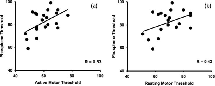
Figure 1.
(a) Relationship between phosphene thresholds (PT) and active motor thresholds (rMT). PT and
aMT are significantly correlated (P ¼ 0.014). (b) Relationship between PT and resting motor
thresholds (rMT) (P ¼ 0.052). All thresholds are in percent of maximum stimulator output.
Subjects that did not perceive phosphenes revealed a 2003], the present data suggest that aMT may help guide slightly higher aMT and rMT (aMT 65.3 6 5.3 SD, rMT stimulation intensity over the visual cortex, and perhaps 74.7 6 8.9) compared with subjects who did perceive phos- over other nonmotor regions. More clearly, as our finding phenes (aMT 59.9 6 8.2 SD; rMT 69.2 6 9.9), but the dif- is at variance with other studies using different methodol- ferences were not significant (aMT, P ¼ 0.14; rMT P ¼ ogies, these data underscore the sensitivity of motor or PT to the details of the thresholding protocol, lighting condi- Repeated measures ANOVA revealed significant differ- tions, and eyes-open or eyes-closed state. This advocates ences between thresholds [F(2,40) ¼ 2642, P < 0.001]. Post- for brain mapping studies that use TMS to provide hoc contrasts revealed significant differences between each detailed statements of procedures, methodology, and TMS pair of thresholds: (a) rMT vs. aMT: mean difference 6 SD factors used when measuring reference thresholds and ¼ 9.33 6 3.98, P < 0.001, (b) rMT vs. PT: 13.76 6 9.59, P < establishing experimental intensities over various brain 0.001, and (c) aMT vs. PT: 22.33 6 9.02, P < 0.0001.
There was a strong positive correlation between rMT The use of different approaches toward threshold (up, and aMT (r ¼ 0.92; P < 0.001). A significant positive corre- down, or other) and different states of visual input (eyes lation was also found between aMT and PT (r ¼ 0.53; P ¼ open or closed/blindfolded), employed in previous studies 0.014; Fig. 1a). A trend toward a correlation was present during MT and PT determinations, may have limited the between rMT and PT (r ¼ 0.43; P ¼ 0.052, Fig. 1b).
ability to find a correlation between the two measures.
Our emphasis on systematic parallel methodology formeasuring MT and PT may have been critical in producing DISCUSSION
All prior studies reported slightly different protocols for In contrast to previous reports [Antal et al., 2003b; Bor- approaching PT. Stewart et al. [2001b] determined PT by oojerdi et al., 2002; Gerwig et al., 2003; Stewart et al., decreasing or increasing TMS intensity in 5% increments 2001b], we found a significant correlation between aMT from a starting point of 60% MSO. Two studies and PT with a trend toward significance between rMT and approached PT from below by increasing intensities in 1 PT. While the aMT and PT correlation was modest, with or 2% increments [Antal et al., 2003b; Boroojerdi et al., aMT accounting for 27.8% of the group variance in PT, our 2002]. Gerwig et al. [2003] established PT by first in- finding is the first to suggest such a relation between TMS creasing intensities by 5% increments to then randomly thresholds in visual and motor cortex. Such a correlation is increasing and decreasing intensities by additional 2% consistent with the idea that there is an element of global excitability specific to each subject if the thresholding pro- In contrast, details about MT procedures were relatively cedures are similar for motor and visual cortex. While it sparse. One study reported approaching MT downward may be useful to determine TMS intensities with relevant by reducing intensities by 2% increments [Stewart et al., region- and task-specific thresholds [Robertson et al., 2001b]. Other studies provided few details about the r Correlation Between Motor and Phosphene Thresholds r
approach to MT determination [Antal et al., 2003b; Boroo- cortex showed greater activation and geniculate nucleus jerdi et al., 2002; Stewart et al., 2001b]. Notably, while a smaller activation under the eyes-open condition, visuomo- downward approach to threshold has been recommended tor structures (e.g., prefrontal and parietal cortices, frontal for MT in recent guidelines [Rothwell et al., 1999], none of eye fields, cerebellar vermis, thalamus, and basal ganglia) the previous studies reported a downward approach to PT revealed greater activation under the eyes-closed condi- [Antal et al., 2003b; Boroojerdi et al., 2002; Gerwig et al., tion. In addition, TMS studies have reported that, after dark adaptation, motor cortex excitability is increased Since both MEPs and phosphene perception show sub- [Leon-Sarmiento et al., 2005], an effect comparable to the stantial trial-to-trial variability, all MT and PT thresholding decreased phosphene threshold after light deprivation procedures in the present and prior studies have used a [Boroojerdi et al., 2001], possibly due to GABAergic and statistical endpoint of at least 50% detection of behavioral glutamatergic mechanisms from corticocortical networks outcome (MEP or phosphene perception) out of 6–10 test connecting motor and visual areas [Bullier et al., 1996; pulses. However, such statistical endpoints may produce Fadiga et al., 2000; Leon-Sarmiento et al., 2005]. Thus, it different thresholds depending on whether it is systemati- seems apparent that the degree of light exposure and hav- cally approached from below or from above. Conse- ing eyes open or closed critically affects both brain activa- quently, it has been proposed to define MT as the mean of tion patterns and cortical excitabilty. However, unlike PT two thresholds, one from above and one from below [Mills [Boroojerdi et al., 2001], the time course of how MT might and Nithi, 1997]. However, this method is time-consuming vary with light deprivation is not known and it is unclear and no more reliable than measuring thresholds by consis- to what degree 15 min of dark-adaptation might have had, tently approaching it downward [Tranulis et al., 2006].
if any, on our MT measurements. Because of the possibility However, MTs obtained with different techniques in the of different time-courses for dark-adaptation effects, we same subject can differ by as much as 8% of MSO [Mills chose to measure MT and PT consistently at 15 and and Nithi, 1997]. Although comparable data regarding 45 minutes respectively after donning goggles rather than differences in PT when approached differently are not randomize the order of MT/PT determinations between available, we explicitly adopted a consistently downward threshold search for both MT and PT.
As expected and consistent with prior literature, we Previous studies investigating MT and PT correlations found significant differences between group mean rMTs, performed PT determinations with either closed eyes aMTs, and PTs. Within each subject, thresholds were con- [Antal et al., 2003b] or blindfolds [Antal et al., 2003b; Bor- sistently highest for PT and lowest for aMT. Because mus- oojerdi et al., 2002; Gerwig et al., 2003; Stewart et al., cle contraction reduces variability of the spinal excitability 2001b]. In contrast, lighting conditions during MT determi- by ensuring suprathreshold activation of spinal motor neu- nation procedures were either done with eyes open rons, similar to other reports, we found that variability of [Stewart et al., 2001b] or not reported [Antal et al., 2003b; aMT was lower than rMT [Antal et al., 2003b; Nitsche Boroojerdi et al., 2002; Gerwig et al., 2003; Stewart et al., et al., 2005]. Voluntary muscle contraction raises motor 2001b]. In either case, it is unlikely that explicit attention cortical excitability (lower aMT), while reducing the spinal was taken to ensure comparable visual exposure and eye- contribution toward variability [Kiers et al., 1993]. In paral- lid state during both MT and PT procedures.
lel, it is possible that raised visual cortical excitability Although PT does not change significantly under brief (lower PT) with dark-adaptation [Boroojerdi et al., 2000a] exposure to different lighting conditions [Kammer and may also reduce the variability of PT which is yet, to our Beck, 2002], longer periods of dark adaptation reduce PT knowledge, to be systematically examined. Thus, we note and increase the yield of TMS induced phosphenes [Boroo- that our correlation was identified between aMT and dark- jerdi et al., 2000a; Marg and Rudiak, 1994]. We determined adapted PT, both of which represent conditions of PT after 45 min of darkness, a time point by which most increased excitability and possibly of reduced variability dark adaptation has taken place [Boroojerdi et al., 2000a].
or measurement noise. Kiers et al. [1993] suggested that Even though dark adaptation may continue beyond this during a relaxed muscle state, changes in cortical excitabil- time point, PT determinations at the same point in time ity in different regions may be relatively independent allows reasonable comparisons between subjects. Since whereas these changes may be positively correlated during time of dark adaptation was not set in most other studies voluntary muscle contraction. By changing tonic levels of comparing MT and PT [Antal et al., 2003b; Boroojerdi variability specific to each modality tested, i.e. for motor et al., 2002; Gerwig et al., 2003; Stewart et al., 2001b], our responses, removing spinal inhibition by muscle contrac- data suggest that aMT may correlate mainly with a suffi- tion; for visual percepts, increasing cortical excitability by dark adaptation, noise in measurements can be reduced.
Even if no light is present, having eyes open or closed Further, aMT and the PT we measured are likely related differentially influences cortical network activation and to the cortical elements or circuitry specific to the coil posi- cortical excitability. An fMRI study demonstrated that acti- tion and orientation used. Differences in magnetic stimula- vation patterns differ between eyes-open and eyes-closed tion parameters between PT and MT procedures also conditions in darkness [Marx et al., 2004]. Whereas visual potentially limit correlations within studies. In one study, r Deblieck et al. r
different stimulator models which generate different wave- excitability: people who see phosphenes were associated form types were used for PT and MT determination [Antal with greater BOLD activation in primary striate and et al., 2003b]. Two studies used paired-pulse TMS over the early extrastriate visual cortex, while those who do not occipital lobe, while single-pulse TMS was used for MT [Antal et al., 2003b; Boroojerdi et al., 2002]. For MT deter- BOLD activation in higher extrastriate areas [Meister et al., minations, coil orientation was consistently oriented per- pendicular to the central sulcus; for PT determinations, the Although we matched many psychophysical aspects of coil handle was held upward in three studies [Antal et al., MT and PT procedures, we recognize that our procedures 2003b; Boroojerdi et al., 2002; Stewart et al., 2001b] and lat- were not purely parallel. MT was objectively quantified by erally in one study [Gerwig et al., 2003]. To control for MEP amplitudes, while PT depended on subjective report- these factors, we employed the same stimulator and ing. While the problem of phosphene perception is by applied single-pulse TMS for all thresholding procedures.
nature a subjective one, future studies might more closely Although recent studies suggest a lateral preference for match MT procedures by asking subjects to report on their current orientation for phosphene induction [Kammer, perception of muscle twitches following TMS over the 1999], we used the cranio-caudal direction used in most motor cortex. Scalp–brain distances also differ over motor previous MT and PT studies [Antal et al., 2003b; Boroo- and visual cortices and without MRI scans, we could not jerdi et al., 2002; Stewart et al., 2001b].
adjust thresholds for scalp–brain distances as has been Whether cortico-cortical or/and cortico-spinal axons are proposed [Stokes et al., 2005]. We did not quantitatively activated in the hand area depend on the orientation of control force or baseline EMG amplitude for aMT, but the TMS-applied magnetic field and the shape of the coil rather monitored baseline EMG qualitatively online, while [Di Lazzaro et al., 2003]. For example, with a biphasic subjects squeezed with a pinch grip estimated at 10–15% stimulator, both biphasic pulses may activate different de- of maximal voluntary force, a level above which further scending volleys depending on the stimulus intensity and MEP facilitation is minimal [Mills, 1999]. While this proce- the direction of current flow. When the current is charac- dure is comparable to some routine assessments of aMT, it terized by a lateromedial direction, such as was the case in also likely introduces greater noise in the aMT measure- this study, both the cortico-spinal and cortico-cortical ment than if controlled. Future studies might control base- axons are activated. Although it has been suggested that line force or EMG more closely during aMT assessment. In the level of muscle contraction does not affect the ampli- addition, follow-up correlational studies that use two fully tude of the D-wave induced by lateromedial magnetic blinded investigators to measure thresholds may help fur- stimulation, the level of excitability of the pyramidal tract ther clarify this issue. With these limitations accounted for, neurons lead to an increase in the size and number of the it is possible our mild correlation between aMT and dark I-wave volleys [Di Lazzaro et al., 2003]. Consequently, the adapted PT could have been further strengthened.
finding that resting and active muscle states may recruit Physiologic differences and similarities exist between different axonal elements may also partially explain why motor and visual cortices that may affect correlations aMT, but not rMT was found correlated to PT. Similarly, between PT and MT. For example, paired-pulse TMS stud- PT is also sensitive to coil orientation with a preference ies over the motor cortex show differential excitatory or for an induced lateromedial current direction [Kammer inhibitory effects dependent on the interstimulus interval et al., 2001a], which suggests that the PT we determined (ISI); in contrast, phosphene detection is facilitated with is specific for our biphasic current and vertical coil orien- paired-pulse TMS over visual cortices, independently of ISI [Sparing et al., 2005]. Pharmacologic studies with TMS Our correlation is limited to those volunteers who saw have shown that MT can be affected by drugs that block phosphenes. We found no significant differences in either voltage-gated sodium and calcium channels (e.g., carbama- aMT or rMT in the six subjects that did not see phos- zepine, phenytoin, and lamotrigine) [Boroojerdi, 2002; phenes versus those who did see phosphenes. While some Chen et al., 1997b; Manganotti et al., 1999; Ziemann et al., studies report that all their subjects discerned phosphenes 1996b], while being unaffected by drugs that block GABA [Boroojerdi et al., 2002; Rauschecker et al., 2004; Stewart receptors (e.g., lorazepam, diazepam, vigabatrin) [Boroo- et al., 2001b], other studies found a similar percentage of jerdi, 2002; Inghilleri et al., 1996; Ziemann et al., 1996a,b].
subjects lacked phosphene perception [Antal et al., In contrast, these drug classes did not affect PT [Boroo- 2003a,b; Boroojerdi et al., 2000b; Meyer et al., 1991; Sparing jerdi, 2002]. These differences continue to underscore the et al., 2005]. It is not certain that subjects who do not see use of region-specific thresholds when possible as refer- phosphenes simply have higher than measurable thresh- olds [Chronicle and Mulleners, 2004]. For example, it may On the other hand, a common excitability across the be differences in cortical anatomy or intrinsic differences brain has been proposed in discussions of neurological dis- in posterior cortical organization that could account some orders with either increased or decreased excitability (e.g., subjects from not perceiving phosphenes. Meister et al.
exemplified by epilepsy as a disorder of hyperexcitability) [2003] proposed that the difference in phosphene percep- [Saugstad, 2005], and some neurophysiologic features of tion may be found in different regions of visual cortical motor and visual cortices have been found to be similar.
r Correlation Between Motor and Phosphene Thresholds r
For example, both MT and PT increase with hyperventila- Artola A, Singer W (1987): Long-term potentiation and NMDA tion [Sparing et al., 2007]; cortical excitability, as measured receptors in rat visual cortex. Nature 330:649–652.
by either MT or PT, is reduced by 1 Hz rTMS over each Bear MF (1996): Progress in understanding NMDA-receptor-de- area [Boroojerdi et al., 2000b; Chen et al., 1997a]; and, pendent synaptic plasticity in the visual cortex. J Physiol(Paris) 90:223–227.
rapid plasticity paradigms such as ischemic deafferentation Boroojerdi B (2002): Pharmacologic influences on TMS effects.
over the motor cortex or dark-adaptation over the visual cortices are both dependent on GABA and NMDA recep- Boroojerdi B, Bushara KO, Corwell B, Immisch I, Battaglia F, tors [Boroojerdi et al., 2001; Buetefisch et al., 2000; Zie- Muellbacher W, Cohen LG (2000a): Enhanced excitability of the mann et al., 2004]. Specifically, some features of synaptic human visual cortex induced by short-term light deprivation.
plasticity [long-term potentiation (LTP) and long-term depression (LTD)], within both primary motor and visual Boroojerdi B, Prager A, Muellbacher W, Cohen LG (2000b): Reduc- cortex appear similar [Bear, 1996; Boroojerdi et al., 2001; tion of human visual cortex excitability using 1 Hz transcranial Buetefisch et al., 2000; Gilbert, 1998; Sanes and Donoghue, magnetic stimulation. Neurology 54:1529–1531.
Boroojerdi B, Battaglia F, Muellbacher W, Cohen LG (2001): Mech- 2000; Ziemann et al., 2004]. In the motor cortex, LTP is anisms underlying rapid experience-dependent plasticity in the dependent on NMDA activation and reduced GABA-ergic human visual cortex. Proc Natl Acad Sci USA 98:14698– inhibition [Hess and Donoghue, 1994; Otsuki et al., 1998], while being unaffected by sodium and calcium channel Boroojerdi B, Meister IG, Foltys H, Sparing R, Cohen LG, Toepper modulation [Buetefisch et al., 2000; Otsuki et al., 1998].
R (2002): Visual and motor cortex excitability: A transcranial Similarly, the direction of GABA-ergic inhibition and magnetic stimulation study. Clin Neurophysiol 113:1501–1504.
NMDA receptor activation, essential for LTP, have been Brasil-Neto JP, Cohen LG, Panizza M, Nilsson J, Roth BJ, Hallett linked with visual plasticity as well [Artola and Singer, M (1992): Optimal focal transcranial magnetic activation of the 1987; Bear, 1996; Quinlan et al., 1999], while being inde- human motor cortex: Effects of coil orientation, shape of the pendent of sodium channel blockade [Boroojerdi et al., induced current pulse, and stimulus intensity. J Clin Neuro-physiol 9:132–136.
2001]. This evidence of shared neurophysiologic features Buetefisch CM, Davis BC, Wise SP, Sawaki L, Kopylev L, Classen between motor and visual cortices may suggest a rationale J, Cohen LG (2000): Mechanisms of use-dependent plasticity in the human motor cortex. Proc Natl Acad Sci USA 97:3661–3665.
Bullier J, Schall JD, Morel A (1996): Functional areas in occipito- CONCLUSION
frontal connections in the monkey. Behav Brain Res 76:89–97.
Chaudry V, Cornblath DR, Mellits ED, Avila O, Freimer ML, Glass Using procedurally similar approaches toward thresh- JD (1991): Inter- and intra-examiner reliability of nerve conduc- olding, we report a significant correlation between aMT tion measurements in normal subjects. Annal Neurol 30:841– over motor cortex and dark-adapted PT over visual cortex.
This correlation provides evidence that a shared, signifi- Chen R, Classen J, Gerloff C, Celnik P, Wassermann EM, Hallett cant global contribution to cortical responsiveness to TMS M, Cohen LG. (1997a): Depression of motor cortex excitability might be present over different cortical regions. Our data by low-frequency transcranial magnetic stimulation. Neurology suggest that TMS thresholds are sensitive to details of thresholding procedure, lighting conditions, and eyes- Chen R, Samii A, Canos M, Wassermann EM, Hallett M. (1997b): Effects of phenytoin on cortical excitability in humans. Neurol- open/eyes-closed state and invite a re-evaluation of meth- ods of threshold determination when comparisons are Chronicle EP, Mulleners WM (2004): Controversies in headache being made across regions. Since aMT are easily measured, [Letter to the Editor]. Cephalalgia 24:317–318.
our data suggest the possibility of using aMT as a guide to Cohen LG, Celnik P, Pascual-Leone A, Corwell B, Faiz L, Dambro- calibrate TMS intensities during TMS mapping studies sia J, Honda M, Sadato N, Gerloff C, Catala MD, Hallett M over the visual cortex and possibly other nonmotor cortical (1997): Functional relevance of cross-modal plasticity in blind regions. In addition, our correlation provides a rationale for current guidelines that calibrate TMS intensity for re- Desmurget M, Epstein CM, Turner RS, Prablanc C, Alexander GE, petitive TMS applications based on MT regardless of corti- Grafton ST (1999): Role of the posterior parietal cortex inupdating reaching movements to a visual target. Nat Neurosci Di Lazzaro V, Oliviero A, Pilato F, Mazzone P, Insola A, Ranieri REFERENCES
F, Tonali PA (2003): Corticospinal volleys evoked by transcra-nial stimulation of the brain in conscious humans. Neurol Res Antal A, Kincses TZ, Nitsche MA, Paulus W (2003a): Manipula- tion of phosphene thresholds by transcranial direct current Fadiga L, Fogassi L, Gallese V, Rizzolatti G (2000): Visuomotor stimulation in man. Exp Brain Res 150:375–378.
neurons: Ambiguity of the discharge or ‘motor’ perception? Int Antal A, Nitsche MA, Kincses TZ, Lampe C, Paulus W (2003b): No correlation between moving phosphene and motor thresh- Gerwig M, Kastrup O, Meyer B-U, Niehaus L (2003): Evaluation olds: A transcranial magnetic stimulation study. Neuroreport of cortical excitibilty by motor and phosphene thresholds in transcranial magnetic stimulation. J Neurol Sci 215:75–78.
r Deblieck et al. r
Gilbert CD (1998): Adult cortical dynamics. Physiol Rev 78:467– Modulating parameters of excitability during and after trans- cranial direct current stimulation of the human motor cortex.
Gothe J, Brandt SA, Irlbacher K, Roericht S, Sabel BA, Meyer BU (2002): Changes in visual cortex excitability in blind subjects as Otsuki K, Morimoto K, Sato K, Yamada N, Kuroda S (1998): demonstrated by transcranial magnetic stimulation. Brain Effects of lamotrigine and conventional antiepileptic drugs on amygdala- and hippocampal-kindled seizures in rats. Epilepsy Heiser M, Iacoboni M, Maeda F, Marcus J, Mazziotta JC (2003): The essential role of Broca’s area in imitation. Eur J Neurosci Quinlan EM, Philpot BD, Huganir RL, Bear MF (1999): Rapid, ex- perience-dependent expression of synaptic NMDA receptors in Hess G, Donoghue JP (1994): Long-term potentiation of horizontal visual cortex in vivo. Natl Neurosci 2:352–357.
connections provides a mechanism to reorganize cortical motor Rauschecker AM, Bestmann S, Walsh V, Thilo KV (2004): Phos- maps. J Neurophysiol 71:2543–2547.
phene threshold as a function of contrast of external visual Hotson JR, Anand S (1999): The selectivity and timing of motion stimuli. Exp Brain Res 157:124–127.
processing in human temporo-parieto-occipital cortex: A trans- Robertson EM, Theoret H, Pascual-Leone A (2003): Studies in cog- cranial magnetic stimulation study. Neuropsychologia 37:169– nition: the problems solved and created by transcranial mag- netic stimulation. J Cogn Neurosci 15:948–960.
Inghilleri M, Berardelli A, Marchetti P, Manfredi M (1996): Effects Rothwell JC, Hallett M, Berardelli A, Eisen A, Rossini PM, Paulus of diazepam, baclofen and thiopental on the silent period W (1999): Magnetic stimulation: motor evoked potentials. The evoked by transcranial magnetic stimulation in humans. Exp International Federation of Clinical Neuropysiology. Electroen- cephalogr Clin Neurophysiol Suppl 52:97–103.
Kammer T (1999): Phosphenes and transient scotomas induced by Sanes JN, Donoghue JP (2000): Plasticity and primary motor cor- magnetic stimulation of the occipital lobe: Their topographic tex. Annu Rev Neurosci 23:393–415.
relationship. Neuropsychologia 37:191–198.
Saugstad LF (2005): The ‘‘new-old’’ way of thinking about brain Kammer T, Beck S (2002): Phosphene thresholds evoked by trans- disorder, cerebral excitability—The fundamental property of cranial magnetic stimulation are insensitive to short-lasting nervous tissue. Med Hypotheses 64:142–492.
variations in ambient light. Exp Brain Res 145:407–410.
Sparing R, Dambeck N, Stock K, Meister IG, Huetter D, Boroojerdi Kammer T, Beck S, Erb M, Grodd W (2001a): The influence of B (2005): Investigation of the primary visual cortex using short- current direction on phosphene thresholds evoked by trans- interval paired-pulse transcranial magnetic stimulation (TMS).
cranial magnetic stimulation. Clin Neurophysiol 112:2015– Sparing R, Dafotakis M, Buelte D, Meister IG, Noth J (2007): Excit- Kiers L, Cros D, Chiappa KH, Fang J (1993): Variability of motor ability of human motor and visual cortex before, during and potentials evoked by transcranial magnetic stimulation. Electro- after hyperventilation. J Appl Physiol 102:405–411.
encephalogr Clin Neurophysiol 89:415–423.
Stewart L, Ellison A, Walsh V, Cowey A. (2001a): The role of Leon-Sarmiento FE, Bara-Jimenez W, Wassermann EM (2005): Vis- transcranial magnetic stimulation (TMS) in studies of vision, ual deprivation effects on human motor cortex excitability.
attention and cognition. Acta Psychol 107:275–291.
Stewart LM, Walsh V, Rothwell JC. (2001b): Motor and phosphene Manganotti P, Bongiovanni LG, Zanette G, Turazzini M, Fiaschi A thresholds: A transcranial magnetic stimulation correlation (1999): Cortical excitability in patients after loading doses of study. Neuropsychologia 39:415–419.
lamotrigine: A study with magnetic brain stimulation. Epilep- Stokes MG, Chambers CD, Gould IC, Henderson TR, Janko NE, Allen NB, Mattingley JB (2005): Simple metric for scaling motor thresh- Marg E, Rudiak D (1994): Phosphenes induced by magnetic stimu- old based on scalp-cortex distance: Application to studies using lation over the occipital brain: description and probable site of transcranial magnetic stimulation. J Neurophysiol 94:4520–4527.
stimulation. Optom Vis Sci 71:301–311.
Tranulis C, Gueguen B, Pham-Scottez A, Vacheron MN, Cabel- Marx E, Deutschlander A, Stephan T, Dieterich M, Wiesmann M, guen G, Costantini A, Valero G, Calinovski A (2006): Motor Brandt T (2004): Eyes open and eyes closed as rest conditions: threshold in transcranial magnetic stimulation: Comparison of Impact on brain activation patterns. Neuroimage 21:1818–1824.
three estimation methods. Neurophysiol Clin 36:1–7.
Meister IG, Weidemann J, Dambeck N, Foltys H, Sparing R, Wassermann EM (1998): Risk and safety of repetitive transcranial Krings T, Thron A, Boroojerdi B (2003): Neural correlates of magnetic stimulation: Report and suggested guidelines from phosphene perception. Suppl Clin Neurophysiol 56:305–311.
the International Workshop on the Safety of Repetitive Trans- Meyer BU, Diehl RR, Steinmetz H, Britton TC, Benecke R (1991): cranial Magnetic Stimulation, June 5–7, 1996. Electroencepha- Magnetic stimuli applied over motor cortex and visual cortex: Influence of coil position and field polarity on motor responses, Ziemann U, Lonnecker S, Steinhoff BJ, Paulus W (1996a): The phosphenes and eye movements. Electroencephalogr Clin Neu- effect of lorazepam on the motor cortical excitability in man.
Mills KR (1999). Magnetic Stimulation of the Human Nervous Sys- Ziemann U, Lonnecker S, Steinhoff BJ, Paulus W (1996b): Effects of tem. New York: Oxford University Press.
antiepileptic drugs on motor cortex excitability in humans: A Mills KR, Nithi K (1997): Corticomotor threshold to magnetic stim- transcranial magnetic stimulation study. Ann Neurol 40:367–378.
ulation: Normal values and repeatability. Muscle Nerve 20: Ziemann U, Iliac TV, Pauli C, Meintzschel F, Ruge D (2004): Learning modifies subsequent induction of long-term potentia- Nitsche MA, Seeber A, Frommann K, Klein CC, Rochford C, Nit- tion-like and long-term depression-like plasticity in human sche MS, Fricke K, Liebetanz D, Lang N, Antal A, et al. (2005): motor cortex. J Neurosci 24:1666–1672.

Source: http://iacoboni.bol.ucla.edu/pdfs/HBM_Deblieck_v29p662.pdf

Protocolli di idoneità.pdf

COMITATO TECNICO-DIRETTIVO DMTE DELLA PROVINCIA DI CREMONA PROTOCOLLI OPERATIVI PER L’ ACCERTAMENTO DELL’ IDONEITA’ DEL DONATORE DI SANGUE E DI EMOCOMPONENTI E LA VALIDAZIONE DELLE UNITA’ RACCOLTE Premesse • Le Strutture Trasfusionali e le Associazioni di volontariato collaborano per mettere a disposizione di tutti i candidati donatori materiale educativo

Halitosis

containing amino acids such as methionine,cysteine and cystine. The resulting volatilereported complaint. Whether in the form ofoccasional ‘morning breath’, which nearlyevery otherwise healthy adult encounters,dimethyl sulphide and dimethyl disulphide)or rarer and more serious problems rangingfrom metabolic disorders to chest tumours,putrescine, foul-smelling diamines) are athalitosis

Copyright © 2011-2018 Health Abstracts