Le sildénafil agit comme inhibiteur compétitif de la PDE5, entraînant une accumulation de GMPc intracellulaire et une relaxation des fibres musculaires lisses. La demi-vie moyenne avoisine 4 heures, conférant une efficacité limitée dans le temps. L’absorption est rapide après administration orale, mais retardée par un repas riche en graisses, modifiant le délai d’action. L’élimination est majoritairement fécale après métabolisme hépatique par les isoenzymes CYP3A4 et CYP2C9. Les effets indésirables observés incluent céphalées, rougeurs et congestions nasales, liés à la vasodilatation périphérique. Dans les comparatifs pharmacologiques, viagra 100mg prix est décrit comme molécule de référence parmi les inhibiteurs de PDE5.
Microsoft word - 35rowlands.targeting the hepa.doc
Targeting the Hepatitis C virus ion channel p7 for anti-viral therapy
Stephen Griffin, Dean Clarke, Steve Evans, Alastair Smith, Joachim Jäger,
Introduction Hepatitis C virus (HCV) currently infects over 3 % of the world population and is the major indicator for liver transplant surgery in the west. Acute infection is usually asymptomatic but leads to persistence in the majority of cases causing chronic liver disease. Treatment of the virus is currently limited to the use of type 1 interferon either alone, or in combination with the guanosine analogue ribavirin, and this therapeutic regime is expensive, poorly tolerated, and effective in only 40 % of cases world-wide. Furthermore, resistance to this treatment is common in the viral genotypes found in the west. The search for a vaccine and alternative therapies has been hampered by the current inability to successfully culture the virus in vitro, making the identification of new anti-viral drug targets paramount. The HCV p7 protein is a small hydrophobic protein of 63 amino acids comprised of two trans-membrane alpha helices separated by a short positively charged cytoplasmic loop (Fig. 1). It is predicted to belong to a family of proteins known as viroporins, which homo-oligomerise to form aqueous pores in cellular membranes. Perhaps the best characterised of these proteins is the M2 channel of Influenza A virus: Fig. 1. Computer the target of the first anti-viral drug, Amantadine.
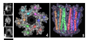
Biophysical studies of p7 oligomers High level expression of a p7 was achieved by fusion with glutathione-S-transferase in E. coli
separated by a 6-histidine linker: GST-HIS-p7. Oligomeric complexes were visualised by Transmission Electron Microscopy (TEM) in collaboration with Dr Lucy Beales (University of Leeds). Both GST-HIS-p7 and cleaved near-native HIS-p7 (Fig. 2) were shown to form ring-like structures with dimensions
Fig. 2. 1-3; TEM of HISp7 oligomers. 4 & 5;
modelling performed by Dr Joachim Jaeger
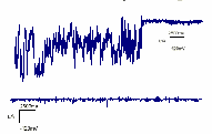
collaborating with the laboratories of Dr Alastair Smith and Professor Jennifer Kirkham (University of Leeds) in order to visualise these complexes by Atomic Force Microscopy (AFM) in lipid membranes under aqueous conditions. In vitro ion channel formation by p7 is inhibited by Amantadine In collaboration with the laboratory of Professor Stephen Evans (University of Leeds), purified HIS-p7 was shown to form ion channels in artificial lipid bilayers using a Black Lipid Membrane (BLM) system. p7 ion channels showed a preference for calcium ions over potassium, consistent with the proteins’ reticular localisation in living Fig. 3. p7 ion channel activity
cells. Furthermore, this activity was abrogated by the before (top) and after (bottom)
addition of Amantadine (Fig. 3). Recent clinical trials that adding 1 µM Amantadine
include this drug with existing therapy show efficacy in many cases, particularly in those patients who were previously non-responsive. We propose that p7 is the target of Amantadine and are working with GlaxoSmithKline to develop new anti-HCV compounds based on this finding. p7 modulates cellular membrane permeability Recently, we have gone on to assess the effect of expressing p7 in the context of living cells. Inducible expression of native p7 in E. coli was shown to cause a growth-inhibitory effect due to membrane leakage, which was reversed by the addition of Amantadine. Furthermore, mutation of the charged loop also negated this effect consistent with studies in the related Pestivirus: Bovine Viral Diarrhoea Virus. We have also demonstrated p7-induced membrane permeability in mammalian cells causing enhanced susceptibility to Hygromycin B. In addition, we have shown in collaboration with Dr Wendy Barclay (University of Reading) that p7 can functionally replace Influenza A M2 protein in a bioassay and is again inhibited by the addition of Amantadine. Further studies of p7 and other viroporins We are currently looking at chimeric viral systems in order to assess the function of p7 in the context of virus replication and it’s ability to functionally complement similar proteins from other viruses. The p7 of the closely related GB Virus B, BVDV p7, Picornavirus 2B (Fig. 4), and Flavivirus NS1 proteins are all candidate systems for such studies. Work has already begun on characterising these proteins and construction of viral chimeras. We are also attempting to crystallise the p7 of HCV and of BVDV, as well as pursuing other structural investigations such as NMR and mass spectroscopy in order to determine the precise composition of the ion channel structure for rational drug design. Fig. 4. GFP-tagged Publication Griffin, S.D., Beales, L.P., Clarke, D.S., Worsfold, O., Evans, S.D., Jaeger, J., Harris, M.P. & Rowlands, D.J. (2003) The p7 protein of hepatitis C virus forms an ion channel that is blocked by the antiviral drug, Amantadine. (2003) FEBS Lett535, 34-8 Funding This work was funded by the MRC “Enabling Technologies” Co-operative, University of Leeds.
Dept. of Computer Science, Faculty of Sciences, Vrije UniversiteitAmsterdam; de Boelelaan 1081a, 1081 HV Amsterdam, The NetherlandsAgents, and in particular mobile agents, offer a means for application developers tobuild distributed applications. Given homogeneity of agent platform and code base, agentmigration is possible. However, many agent platforms exist, differing substantially in thesupp
P E N I N S U L A B I B L E C H U R C H C U P E R T I N Oin our text today, Jesus extends an invitation to his disciples to were lying around hippos like little chicks gathered around accompany him to “the other side of the lake” to expand the sphere the mother hen. Kursi, a fishing village in the northwestern of his mission. This will be Jesus’ first venture into Gentile territory co
 Targeting the Hepatitis C virus ion channel p7 for anti-viral therapy
  include this drug with existing therapy show efficacy in many cases, particularly in those